Line Drawing 1
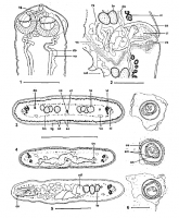
Figs. 1-6. Ophiotaenia oumanskyi sp. n. from Lepidobatrachus laevis. (1) MHNG-PLAT-62560, holotype 1. Scolex, dorsoventral view. (2) MHNG-PLAT-82004, paratype. Cirrus-sac and vagina, dorsal view; note... MoreFigs. 1-6. Ophiotaenia oumanskyi sp. n. from Lepidobatrachus laevis. (1) MHNG-PLAT-62560, holotype 1. Scolex, dorsoventral view. (2) MHNG-PLAT-82004, paratype. Cirrus-sac and vagina, dorsal view; note the presence of a vaginal sphincter. (3) MHNG-PLAT-82005, paratype. Mature proglottis, transverse section of posterior part level. (4) MHNG-PLAT-82005, paratype. Mature proglottis, transverse section at ovarian level. (5) Cross-section of gravid proglottis, at level of anterior part (6) MHNG-PLAT-82005, paratype 2. Eggs drawn in distilled water. Abbreviations: cg = glandular cells, probably of exocrine type, ci = cirrus, cs = cirrus-sac, do = dorsal osmoregulatory canal, em = embryophore, lm = internal longitudinal musculature, ln = longitudinal lateral nerves, oc = outer envelope, on = oncosphere, ov = ovary, sc = subtegumental cells, st = subtegumental muscle fibres, te = testes, tg = tegument, ud = uterine diverticula, ut = uterus, va = vas deferens, vc = vaginal canal, vi = vitelline follicles, vo = ventral osmoregulatory canal, vs = vaginal sphincter. Scale bars: 1, 5 = 250 µm; 2 = 100 µm, 3-4 = 500 µm, 6 = 20 µm. |
Line Drawing 2

Figs 7, 8. Ophiotaenia oumanskyi sp. n. from Lepidobatrachus laevis. (7) MHNG-PLAT-62560, holotype, mature proglottis, dorsal view. (8) MHNG-PLAT-82004, paratype. Gravid proglottis, ventral view. Scal... MoreFigs 7, 8. Ophiotaenia oumanskyi sp. n. from Lepidobatrachus laevis. (7) MHNG-PLAT-62560, holotype, mature proglottis, dorsal view. (8) MHNG-PLAT-82004, paratype. Gravid proglottis, ventral view. Scale-bars: 7, 8 = 500 µm. |
Photo Micrograph
|
Scanning Electron Micrograph
|
 Figs. 1-6. Ophiotaenia oumanskyi sp. n. from Lepidobatrachus laevis. (1) MHNG-PLAT-62560, holotype 1. Scolex, dorsoventral view. (2) MHNG-PLAT-82004, paratype. Cirrus-sac and vagina, dorsal view; note the presence of a vaginal sphincter. (3) MHNG-PLAT-82005, paratype. Mature proglottis, transverse section of posterior part level. (4) MHNG-PLAT-82005, paratype. Mature proglottis, transverse section at ovarian level. (5) Cross-section of gravid proglottis, at level of anterior part (6) MHNG-PLAT-82005, paratype 2. Eggs drawn in distilled water. Abbreviations: cg = glandular cells, probably of exocrine type, ci = cirrus, cs = cirrus-sac, do = dorsal osmoregulatory canal, em = embryophore, lm = internal longitudinal musculature, ln = longitudinal lateral nerves, oc = outer envelope, on = oncosphere, ov = ovary, sc = subtegumental cells, st = subtegumental muscle fibres, te = testes, tg = tegument, ud = uterine diverticula, ut = uterus, va = vas deferens, vc = vaginal canal, vi = vitelline follicles, vo = ventral osmoregulatory canal, vs = vaginal sphincter. Scale bars: 1, 5 = 250 µm; 2 = 100 µm, 3-4 = 500 µm, 6 = 20 µm.
 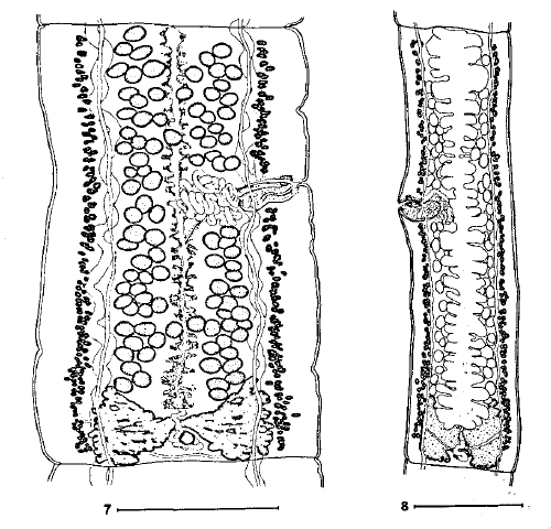 Figs 7, 8. Ophiotaenia oumanskyi sp. n. from Lepidobatrachus laevis. (7) MHNG-PLAT-62560, holotype, mature proglottis, dorsal view. (8) MHNG-PLAT-82004, paratype. Gravid proglottis, ventral view. Scale-bars: 7, 8 = 500 µm.
 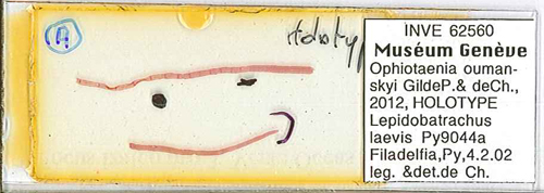 Holotype: MNHG-INVE No. 62560
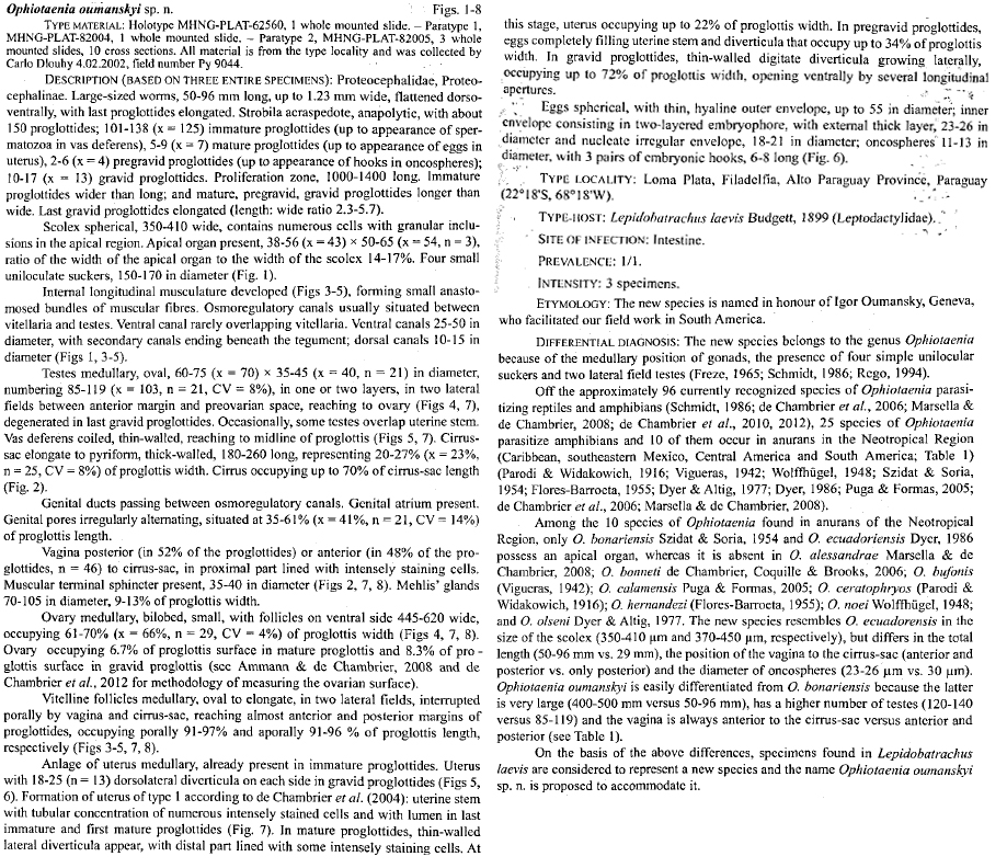

