Line Drawing 1
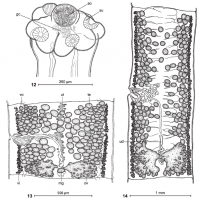
FIGURES 1214. Pangasiocestus romani n. gen. and n. sp. (VNT 298x; holotype: MHNG INVE 79163) from P. larnaudii, Cambodia, line drawings. (12) Scolex, dorsal view. (13) Immature proglottis, dorsal vie... MoreFIGURES 1214. Pangasiocestus romani n. gen. and n. sp. (VNT 298x; holotype: MHNG INVE 79163) from P. larnaudii, Cambodia, line drawings. (12) Scolex, dorsal view. (13) Immature proglottis, dorsal view. (14) Gravid proglottis, dorsal. Abbreviations: ao: apical organ; gc: gland cells; mg: Mehlis gland; ov: ovary; su: sucker; te: testes; ud: uteroduct; ut: uterus; vi: vitelline follicles; vo: ventral osmoregulatory canal. |
Line Drawing 2
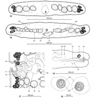
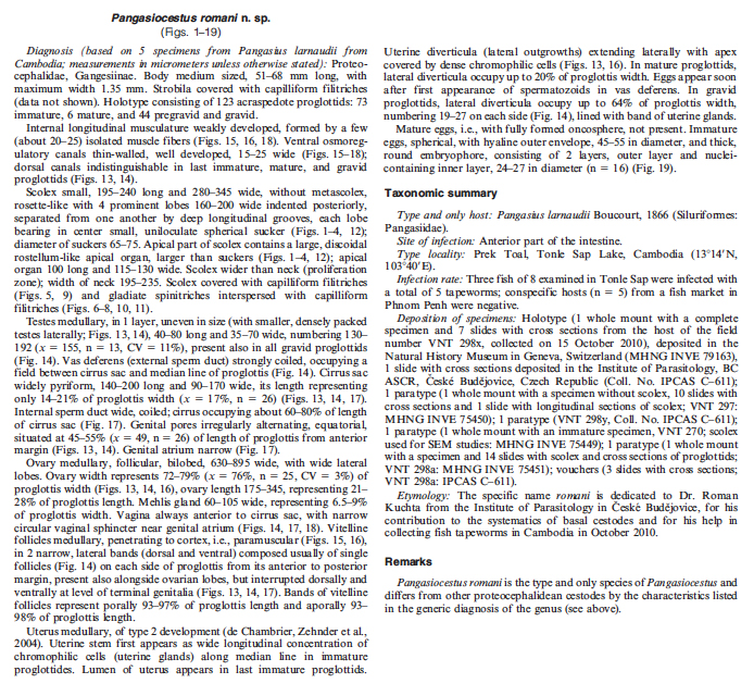
FIGURES 1519. Pangasiocestus romani n. gen., and n. sp. from P. larnaudii, Cambodia, line drawings. (15) Cross section at level of the anterior part
of proglottis (VNT 297; paratype: MHNG INVE 75450... MoreFIGURES 1519. Pangasiocestus romani n. gen., and n. sp. from P. larnaudii, Cambodia, line drawings. (15) Cross section at level of the anterior part
of proglottis (VNT 297; paratype: MHNG INVE 75450). (16) Cross section at level of the ovary (VNT 298x; holotype: MHNG INVE 79163). (17)
Terminal genitalia, dorsal (VNT 298y; paratype: IPCAS C611). (18) Cross section at level of the vagina, showing the vaginal sphincter (VNT 298x;
holotype: MHNG INVE 79163). (19) Immature eggs (VNT 298y; paratype: IPCAS C611). Abbreviations: cc: chromophilic cells; ci: cirrus; cs: cirrus sac; do: dorsal osmoregulatory canal; du: uterine diverticula; em: bi-layered embryophore; lm: internal longitudinal musculature; oe: outer envelope; ov:
ovary; te: testes; ut: uterus; va: vas deferens; vc: vaginal canal; vi: vitelline follicles; vo: ventral osmoregulatory canal; vs: vaginal sphincter. |
Photo Micrograph
|
Scanning Electron Micrograph
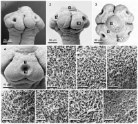
FIGURES 111. Pangasiocestus romani n. gen. and n. sp. from Pangasius larnaudii, Cambodia (VNT 270; paratype: MHNG INVE 75449). Scanning electron micrographs. (1) Scolex, dorsoventral. (2) Scolex, lat... MoreFIGURES 111. Pangasiocestus romani n. gen. and n. sp. from Pangasius larnaudii, Cambodia (VNT 270; paratype: MHNG INVE 75449). Scanning electron micrographs. (1) Scolex, dorsoventral. (2) Scolex, lateral. (3) Scolex, apical. (4) Detail of a sucker. (5) Capilliform filitriches near the center of the
apical organ. (6) Gladiate spinitriches interspersed with capilliform filitriches on the surface of the middle part of the pyramidal apex. (7) Gladiate spinitriches interspersed with capilliform filitriches on the external upper rim of suckers. (8) Gladiate spinitriches interspersed with capilliform filitriches on the internal lateral surface of suckers. (9) Capilliform filitriches on the external upper surface of suckers. (10) Gladiate spinitriches interspersed with
capilliform filitriches on the prominent lobe surrounding the sucker. (11) Gladiate spinitriches interspersed with capilliform filitriches on the internal central surface of suckers. |
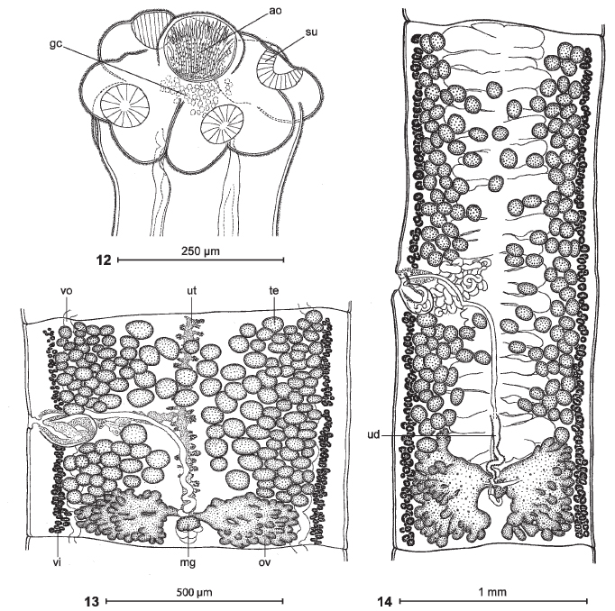 FIGURES 1214. Pangasiocestus romani n. gen. and n. sp. (VNT 298x; holotype: MHNG INVE 79163) from P. larnaudii, Cambodia, line drawings. (12) Scolex, dorsal view. (13) Immature proglottis, dorsal view. (14) Gravid proglottis, dorsal. Abbreviations: ao: apical organ; gc: gland cells; mg: Mehlis gland; ov: ovary; su: sucker; te: testes; ud: uteroduct; ut: uterus; vi: vitelline follicles; vo: ventral osmoregulatory canal.
  FIGURES 1519. Pangasiocestus romani n. gen., and n. sp. from P. larnaudii, Cambodia, line drawings. (15) Cross section at level of the anterior part
of proglottis (VNT 297; paratype: MHNG INVE 75450). (16) Cross section at level of the ovary (VNT 298x; holotype: MHNG INVE 79163). (17)
Terminal genitalia, dorsal (VNT 298y; paratype: IPCAS C611). (18) Cross section at level of the vagina, showing the vaginal sphincter (VNT 298x;
holotype: MHNG INVE 79163). (19) Immature eggs (VNT 298y; paratype: IPCAS C611). Abbreviations: cc: chromophilic cells; ci: cirrus; cs: cirrus sac; do: dorsal osmoregulatory canal; du: uterine diverticula; em: bi-layered embryophore; lm: internal longitudinal musculature; oe: outer envelope; ov:
ovary; te: testes; ut: uterus; va: vas deferens; vc: vaginal canal; vi: vitelline follicles; vo: ventral osmoregulatory canal; vs: vaginal sphincter.
FIGURES 1519. Pangasiocestus romani n. gen., and n. sp. from P. larnaudii, Cambodia, line drawings. (15) Cross section at level of the anterior part
of proglottis (VNT 297; paratype: MHNG INVE 75450). (16) Cross section at level of the ovary (VNT 298x; holotype: MHNG INVE 79163). (17)
Terminal genitalia, dorsal (VNT 298y; paratype: IPCAS C611). (18) Cross section at level of the vagina, showing the vaginal sphincter (VNT 298x;
holotype: MHNG INVE 79163). (19) Immature eggs (VNT 298y; paratype: IPCAS C611). Abbreviations: cc: chromophilic cells; ci: cirrus; cs: cirrus sac; do: dorsal osmoregulatory canal; du: uterine diverticula; em: bi-layered embryophore; lm: internal longitudinal musculature; oe: outer envelope; ov:
ovary; te: testes; ut: uterus; va: vas deferens; vc: vaginal canal; vi: vitelline follicles; vo: ventral osmoregulatory canal; vs: vaginal sphincter. 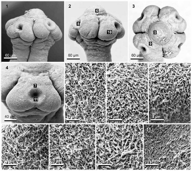 FIGURES 111. Pangasiocestus romani n. gen. and n. sp. from Pangasius larnaudii, Cambodia (VNT 270; paratype: MHNG INVE 75449). Scanning electron micrographs. (1) Scolex, dorsoventral. (2) Scolex, lateral. (3) Scolex, apical. (4) Detail of a sucker. (5) Capilliform filitriches near the center of the
apical organ. (6) Gladiate spinitriches interspersed with capilliform filitriches on the surface of the middle part of the pyramidal apex. (7) Gladiate spinitriches interspersed with capilliform filitriches on the external upper rim of suckers. (8) Gladiate spinitriches interspersed with capilliform filitriches on the internal lateral surface of suckers. (9) Capilliform filitriches on the external upper surface of suckers. (10) Gladiate spinitriches interspersed with
capilliform filitriches on the prominent lobe surrounding the sucker. (11) Gladiate spinitriches interspersed with capilliform filitriches on the internal central surface of suckers.
FIGURES 111. Pangasiocestus romani n. gen. and n. sp. from Pangasius larnaudii, Cambodia (VNT 270; paratype: MHNG INVE 75449). Scanning electron micrographs. (1) Scolex, dorsoventral. (2) Scolex, lateral. (3) Scolex, apical. (4) Detail of a sucker. (5) Capilliform filitriches near the center of the
apical organ. (6) Gladiate spinitriches interspersed with capilliform filitriches on the surface of the middle part of the pyramidal apex. (7) Gladiate spinitriches interspersed with capilliform filitriches on the external upper rim of suckers. (8) Gladiate spinitriches interspersed with capilliform filitriches on the internal lateral surface of suckers. (9) Capilliform filitriches on the external upper surface of suckers. (10) Gladiate spinitriches interspersed with
capilliform filitriches on the prominent lobe surrounding the sucker. (11) Gladiate spinitriches interspersed with capilliform filitriches on the internal central surface of suckers.  Holotype: MNHG-INVE No. 79163

