Line Drawing 1
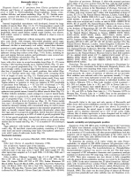
Figures 7-10. Barsonella lafoni n. gen., n. sp. (7) Dorsoventral view of scolex (IPCAS C-485), (8) Sublateral view of scolex (INVE 60358), specimens from Clarias cf. anguillaris, Nile, Nubia Lake rese... MoreFigures 7-10. Barsonella lafoni n. gen., n. sp. (7) Dorsoventral view of scolex (IPCAS C-485), (8) Sublateral view of scolex (INVE 60358), specimens from Clarias cf. anguillaris, Nile, Nubia Lake reservoir. (9,10) Longitudinal secion of solices; specimens from Clarias gariepinus, Tana Lake, Ethiopia (INVE 60353 and 60354, respectively). Abbreviations: ad, additional opening; ao, apical organ; cm, circular musculature. Scale bars: 7-9 = 500 µm; 10 = 250 µm. |
Line Drawing 2

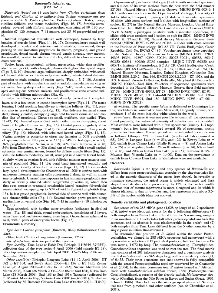
Figures 11-13. Barsonella lafoni n. gen., n. sp. from Clarias gariepinus, Tana Lake, Ethiopia. (11) Holotype, mature proglottid, ventral view (INVE 60346). (12) Holotype, gravid proglottid, dorsal vie... MoreFigures 11-13. Barsonella lafoni n. gen., n. sp. from Clarias gariepinus, Tana Lake, Ethiopia. (11) Holotype, mature proglottid, ventral view (INVE 60346). (12) Holotype, gravid proglottid, dorsal view (INVE 60346). (13) Paratype, sketch of gravid proglottid with 10 uterine pores (up), ventral view (INVE 49395). Scale bars: 11 = 500 µm; 12, 13 = 1 mm
Figures 14-18. Barsonella lafoni n. gen., n. sp. (14) Cross section through uterine pore; specimen from Clarias gariepinus, Langano Lake, Ethiopia (INVE 49398). (15) Terminal genitalia, dorsal view; specimen from C. gariepinus, Langano Lake, Ethiopia (INVE 60359). (16, 17) Cross sections at level of ovary and uterine pore, respectively; specimens from Clarias cf. anguillaris, Nile, Reservoir to Nubia Lake (INVE 60360 and 60352). (18) Eggs in distilled water, with collapsed outer hyaline enevlope (INVE 60361). Abbreviations: cc, chromophilic cells; cs, cirrus sac; em = bilayered embryophore; lm, longitudinal internal musculature; ln, longitudinal nerve cord; mic, microtriches; oe, outer envelope; on, oncospheres; ov, ovary; te, testes; up, uterine pore; ut, uterus; va, vagina; vi, vitelline follicles; vs, vaginal sphincter. Scale bars: 14, 15 = 250 µm; 16, 17 = 1 mm; 18 = 20 µm. |
Photo Micrograph
|
Scanning Electron Micrograph

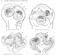
Figures 1-6. Scanning electron micrographs of scolex of Barsonella lafoni n. gen., n. sp. (1,2) Dorsovetral and apical views, respectively; specimen from Clarias gariepinus, Tana Lake, Ethiopia (INVE... MoreFigures 1-6. Scanning electron micrographs of scolex of Barsonella lafoni n. gen., n. sp. (1,2) Dorsovetral and apical views, respectively; specimen from Clarias gariepinus, Tana Lake, Ethiopia (INVE 60351). (3,5) Dorsoventral and apical views, respectively; specimen from Clarias cf. anguillaris, Nubia Lake Reservoir, Sudan (INVE 60352). Black arrows show additional sucker opening (Figs. 1-3). (4) Detail of additional sucker opening; specimen from C. cf. anguillarais, Nubia Lake Reservoir, Sudan (INVE 60350). (6) Microtriches on scolex apex (INVE 60351). Scale bars: 1-3, 5 = 100 µm; 4 = 20 µm; 6 = 2 µm. |
 Figures 7-10. Barsonella lafoni n. gen., n. sp. (7) Dorsoventral view of scolex (IPCAS C-485), (8) Sublateral view of scolex (INVE 60358), specimens from Clarias cf. anguillaris, Nile, Nubia Lake reservoir. (9,10) Longitudinal secion of solices; specimens from Clarias gariepinus, Tana Lake, Ethiopia (INVE 60353 and 60354, respectively). Abbreviations: ad, additional opening; ao, apical organ; cm, circular musculature. Scale bars: 7-9 = 500 µm; 10 = 250 µm.
 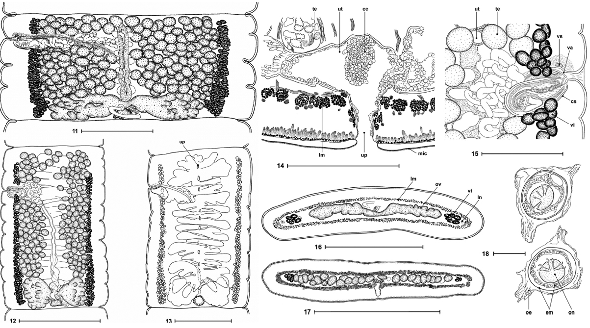 Figures 11-13. Barsonella lafoni n. gen., n. sp. from Clarias gariepinus, Tana Lake, Ethiopia. (11) Holotype, mature proglottid, ventral view (INVE 60346). (12) Holotype, gravid proglottid, dorsal view (INVE 60346). (13) Paratype, sketch of gravid proglottid with 10 uterine pores (up), ventral view (INVE 49395). Scale bars: 11 = 500 µm; 12, 13 = 1 mm
Figures 14-18. Barsonella lafoni n. gen., n. sp. (14) Cross section through uterine pore; specimen from Clarias gariepinus, Langano Lake, Ethiopia (INVE 49398). (15) Terminal genitalia, dorsal view; specimen from C. gariepinus, Langano Lake, Ethiopia (INVE 60359). (16, 17) Cross sections at level of ovary and uterine pore, respectively; specimens from Clarias cf. anguillaris, Nile, Reservoir to Nubia Lake (INVE 60360 and 60352). (18) Eggs in distilled water, with collapsed outer hyaline enevlope (INVE 60361). Abbreviations: cc, chromophilic cells; cs, cirrus sac; em = bilayered embryophore; lm, longitudinal internal musculature; ln, longitudinal nerve cord; mic, microtriches; oe, outer envelope; on, oncospheres; ov, ovary; te, testes; up, uterine pore; ut, uterus; va, vagina; vi, vitelline follicles; vs, vaginal sphincter. Scale bars: 14, 15 = 250 µm; 16, 17 = 1 mm; 18 = 20 µm.
Figures 11-13. Barsonella lafoni n. gen., n. sp. from Clarias gariepinus, Tana Lake, Ethiopia. (11) Holotype, mature proglottid, ventral view (INVE 60346). (12) Holotype, gravid proglottid, dorsal view (INVE 60346). (13) Paratype, sketch of gravid proglottid with 10 uterine pores (up), ventral view (INVE 49395). Scale bars: 11 = 500 µm; 12, 13 = 1 mm
Figures 14-18. Barsonella lafoni n. gen., n. sp. (14) Cross section through uterine pore; specimen from Clarias gariepinus, Langano Lake, Ethiopia (INVE 49398). (15) Terminal genitalia, dorsal view; specimen from C. gariepinus, Langano Lake, Ethiopia (INVE 60359). (16, 17) Cross sections at level of ovary and uterine pore, respectively; specimens from Clarias cf. anguillaris, Nile, Reservoir to Nubia Lake (INVE 60360 and 60352). (18) Eggs in distilled water, with collapsed outer hyaline enevlope (INVE 60361). Abbreviations: cc, chromophilic cells; cs, cirrus sac; em = bilayered embryophore; lm, longitudinal internal musculature; ln, longitudinal nerve cord; mic, microtriches; oe, outer envelope; on, oncospheres; ov, ovary; te, testes; up, uterine pore; ut, uterus; va, vagina; vi, vitelline follicles; vs, vaginal sphincter. Scale bars: 14, 15 = 250 µm; 16, 17 = 1 mm; 18 = 20 µm. 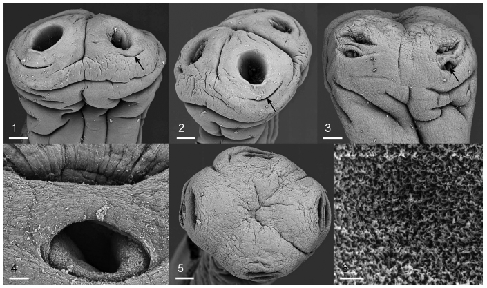 Figures 1-6. Scanning electron micrographs of scolex of Barsonella lafoni n. gen., n. sp. (1,2) Dorsovetral and apical views, respectively; specimen from Clarias gariepinus, Tana Lake, Ethiopia (INVE 60351). (3,5) Dorsoventral and apical views, respectively; specimen from Clarias cf. anguillaris, Nubia Lake Reservoir, Sudan (INVE 60352). Black arrows show additional sucker opening (Figs. 1-3). (4) Detail of additional sucker opening; specimen from C. cf. anguillarais, Nubia Lake Reservoir, Sudan (INVE 60350). (6) Microtriches on scolex apex (INVE 60351). Scale bars: 1-3, 5 = 100 µm; 4 = 20 µm; 6 = 2 µm.
  Holotype: MNHG-INVE No. 60346

