Line Drawing 1
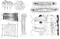
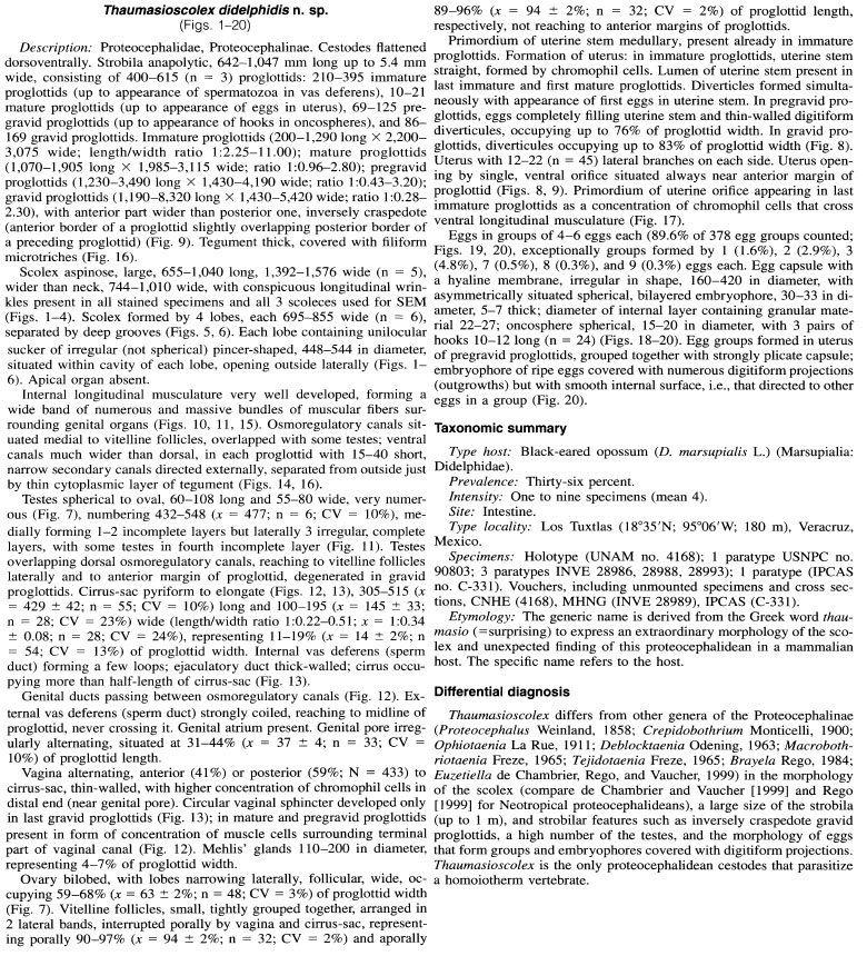
FIGURES1 0-14. Thaumasioscolex didelphidis n. sp. 10, 11. Paratype INVE 28986, cross sections of pregravid proglottids at level of ovary
(10, semithin section) and uterus (11). 12-14. Holotype, UNAM ... MoreFIGURES1 0-14. Thaumasioscolex didelphidis n. sp. 10, 11. Paratype INVE 28986, cross sections of pregravid proglottids at level of ovary
(10, semithin section) and uterus (11). 12-14. Holotype, UNAM 4168, cirrus-sac and vagina (12, dorsal view; 13, ventral view) and 14, detail of
ventral osmoregulatory canal with numerous secondary canals directed to ventral surface. Abbreviations: cv, vaginal canal; dc, dorsal osmoregulatory
canal; lm, longitudinal internal musculature; ov, ovary; sc, secondary osmoregulatory canals; te, testes; ut, uterus; vc, ventral osmoregulatory
canal; vd, vitelline duct; vs, vaginal sphincter; vt, vitelline follicles. Scale bars, 10, 11 = 1,000 µm; 12-14 = 500 µm. |
Line Drawing 2
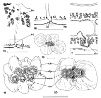
FIGURES1 5-20. Thaumasioscolex didelphidis n. sp. 15-17. Paratype INVE 28986, cross sections, details of secondary canals covered with
thin cytoplasmatic layer of tegument in pregravid proglottids (1... MoreFIGURES1 5-20. Thaumasioscolex didelphidis n. sp. 15-17. Paratype INVE 28986, cross sections, details of secondary canals covered with
thin cytoplasmatic layer of tegument in pregravid proglottids (15, 16) (16, enlargment of 15); cross section of pregravid proglottid with preformed
ventral uterine orifice (17). 18. Holotype, UNAM 4168, eggs spontaneously released to water. 19, 20. Paratype INVE 28986, eggs fixed with
formaldehyde solution and mounted in distilled water (19, upper view; 20, lateral view; note digitiform projections on external surface of egg
embryophore). Abbreviations: ca, capsule; cl, cytoplasmic layer of tegument; dp, digitiform projections on embryophore; em, embryophore; lm,
longitudinal internal musculature; mi, microtriches; on, oncosphere; sc, secondary osmoregulatory canals; uo, uterine orifice. Scale bars, 15, 18-20
= 100 µm; 16 = 50 µm; 17 = 250 µm. |
Photo Micrograph
|
Scanning Electron Micrograph
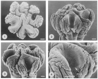
FIGURES1 -4. Thaumasioscolex didelphidis n. sp., paratype INVE 28993, SEM photomicrographs of scolex. 1. Apical view. 2. Lateral view.
3. Subapical view. 4. Detail of lobe with nonspherical sucker (a... MoreFIGURES1 -4. Thaumasioscolex didelphidis n. sp., paratype INVE 28993, SEM photomicrographs of scolex. 1. Apical view. 2. Lateral view.
3. Subapical view. 4. Detail of lobe with nonspherical sucker (arrow showing the margin of the sucker = ms). Abbreviation: ms, margin of
suckers. Scale bars = 100 µm. |
 FIGURES1 0-14. Thaumasioscolex didelphidis n. sp. 10, 11. Paratype INVE 28986, cross sections of pregravid proglottids at level of ovary
(10, semithin section) and uterus (11). 12-14. Holotype, UNAM 4168, cirrus-sac and vagina (12, dorsal view; 13, ventral view) and 14, detail of
ventral osmoregulatory canal with numerous secondary canals directed to ventral surface. Abbreviations: cv, vaginal canal; dc, dorsal osmoregulatory
canal; lm, longitudinal internal musculature; ov, ovary; sc, secondary osmoregulatory canals; te, testes; ut, uterus; vc, ventral osmoregulatory
canal; vd, vitelline duct; vs, vaginal sphincter; vt, vitelline follicles. Scale bars, 10, 11 = 1,000 µm; 12-14 = 500 µm.
FIGURES1 0-14. Thaumasioscolex didelphidis n. sp. 10, 11. Paratype INVE 28986, cross sections of pregravid proglottids at level of ovary
(10, semithin section) and uterus (11). 12-14. Holotype, UNAM 4168, cirrus-sac and vagina (12, dorsal view; 13, ventral view) and 14, detail of
ventral osmoregulatory canal with numerous secondary canals directed to ventral surface. Abbreviations: cv, vaginal canal; dc, dorsal osmoregulatory
canal; lm, longitudinal internal musculature; ov, ovary; sc, secondary osmoregulatory canals; te, testes; ut, uterus; vc, ventral osmoregulatory
canal; vd, vitelline duct; vs, vaginal sphincter; vt, vitelline follicles. Scale bars, 10, 11 = 1,000 µm; 12-14 = 500 µm.  FIGURES1 5-20. Thaumasioscolex didelphidis n. sp. 15-17. Paratype INVE 28986, cross sections, details of secondary canals covered with
thin cytoplasmatic layer of tegument in pregravid proglottids (15, 16) (16, enlargment of 15); cross section of pregravid proglottid with preformed
ventral uterine orifice (17). 18. Holotype, UNAM 4168, eggs spontaneously released to water. 19, 20. Paratype INVE 28986, eggs fixed with
formaldehyde solution and mounted in distilled water (19, upper view; 20, lateral view; note digitiform projections on external surface of egg
embryophore). Abbreviations: ca, capsule; cl, cytoplasmic layer of tegument; dp, digitiform projections on embryophore; em, embryophore; lm,
longitudinal internal musculature; mi, microtriches; on, oncosphere; sc, secondary osmoregulatory canals; uo, uterine orifice. Scale bars, 15, 18-20
= 100 µm; 16 = 50 µm; 17 = 250 µm.
FIGURES1 5-20. Thaumasioscolex didelphidis n. sp. 15-17. Paratype INVE 28986, cross sections, details of secondary canals covered with
thin cytoplasmatic layer of tegument in pregravid proglottids (15, 16) (16, enlargment of 15); cross section of pregravid proglottid with preformed
ventral uterine orifice (17). 18. Holotype, UNAM 4168, eggs spontaneously released to water. 19, 20. Paratype INVE 28986, eggs fixed with
formaldehyde solution and mounted in distilled water (19, upper view; 20, lateral view; note digitiform projections on external surface of egg
embryophore). Abbreviations: ca, capsule; cl, cytoplasmic layer of tegument; dp, digitiform projections on embryophore; em, embryophore; lm,
longitudinal internal musculature; mi, microtriches; on, oncosphere; sc, secondary osmoregulatory canals; uo, uterine orifice. Scale bars, 15, 18-20
= 100 µm; 16 = 50 µm; 17 = 250 µm.  FIGURES1 -4. Thaumasioscolex didelphidis n. sp., paratype INVE 28993, SEM photomicrographs of scolex. 1. Apical view. 2. Lateral view.
3. Subapical view. 4. Detail of lobe with nonspherical sucker (arrow showing the margin of the sucker = ms). Abbreviation: ms, margin of
suckers. Scale bars = 100 µm.
FIGURES1 -4. Thaumasioscolex didelphidis n. sp., paratype INVE 28993, SEM photomicrographs of scolex. 1. Apical view. 2. Lateral view.
3. Subapical view. 4. Detail of lobe with nonspherical sucker (arrow showing the margin of the sucker = ms). Abbreviation: ms, margin of
suckers. Scale bars = 100 µm. 

