Cestode Scientific Name
| Species ID | 9802 |
|---|---|
| Order | Phyllobothriidea |
| Family | Phyllobothriidae |
| Subfamily | |
| Genus | Hemipristicola |
| Species | gunterae |
| Authority | Cutmore, Theiss, Bennett and Cribb, 2011 |
| Taxonomic Status | Valid |
| Valid Name | |
| Synonyms | |
| Genus Record | No |
| Type Species | Yes |
| Verified | No |
| Verified By | |
| Citation(s) |
Cutmore, S. C., S. M. Theiss, M. B. Bennett, and T.H. Cribb. 2011. Hemipristicola gunterae gen. n., sp. n. (Cestoda: Tetraphyllidea: Phyllobothriidae) from the snaggletooth shark, Hemipristis elongata (Carcharhiniformes: Hemigaleidae), from Moreton Bay, Australia. Folia Parasitologica 58: 187-196. (5905) Download PDF |
| Redescription | |
| Scientific Name Notes |
Record Data
| Date (MM/DD/YYYY) | Action | User Name |
|---|---|---|
| 08/03/2012 | Created | T.R. Ruhnke |
| 06/06/2015 | Modified | T. Ruhnke |
Type Host
| Host Class | |||||||
|---|---|---|---|---|---|---|---|
| Host Order | Carcharhiniformes | ||||||
| Host Family | Hemigaleidae | ||||||
|
Type Host (Literal) |
|
||||||
|
Type Host (Valid) |
|
||||||
| Additional Host(s) | |||||||
| Site in Host | |||||||
| Host Notes |
Type Locality
| Country | Australia |
|---|---|
| Body of Water | Moreton Bay |
| Island(s) | |
| City/Region | Wynnum banks |
| Coordinates | |
| DD Latitude | -27.416 |
| DD Longitude | 153.166 |
| Additional Localities | |
| Locality Notes |
Specimens
| Type Material | Holotype (QM G 232173) and 8 paratypes(QM G 23 217481) |
|---|---|
| Total Number of Type Specimens | 9 |
| Voucher Material | |
| Specimen Notes |
Data are given as in original description unless otherwise indicated.
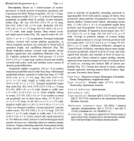


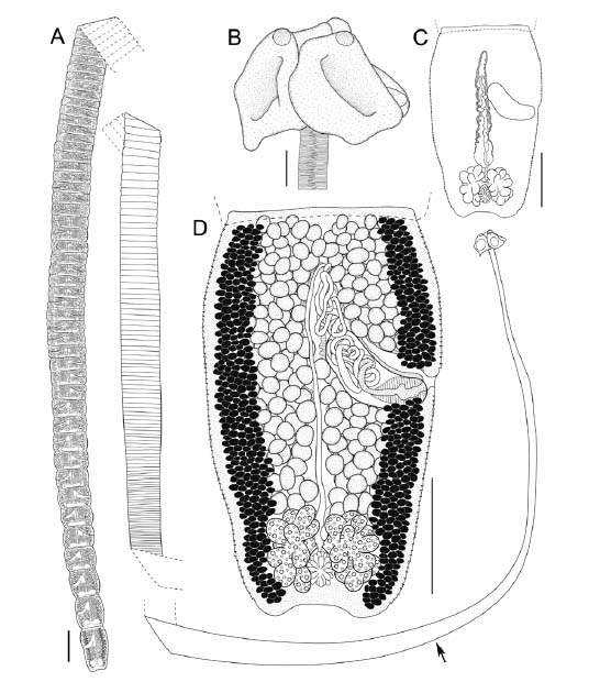 Fig. 1. Hemipristicola gunterae gen. n., sp. n. (holotype QM G 232173), line drawings. A whole worm (black arrow indicates
boundary between neck and strobila); B scolex; C terminal proglottid with testes and vitelline follicles removed to highlight
position of uterus and uterine duct; D terminal proglottid, dorsal view (note: uterine duct not illustrated). Scale bars: A = 1 mm;
B = 200 μm; C, D = 400 μm.
Fig. 1. Hemipristicola gunterae gen. n., sp. n. (holotype QM G 232173), line drawings. A whole worm (black arrow indicates
boundary between neck and strobila); B scolex; C terminal proglottid with testes and vitelline follicles removed to highlight
position of uterus and uterine duct; D terminal proglottid, dorsal view (note: uterine duct not illustrated). Scale bars: A = 1 mm;
B = 200 μm; C, D = 400 μm. 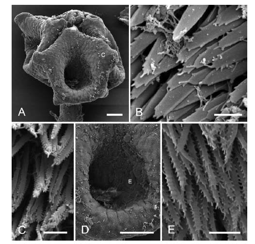 Fig. 2. Hemipristicola gunterae gen. n., sp. n., scanning electron micrographs. A scolex (letter indicates where C was taken);
B proximal bothridial surface; C distal bothridial surface; D bothridial cavity (letter indicates where E was taken); E distal
bothridial surface in centre of bothridial cavity. Scale bars: A, D = 100 μm; B, C, E = 1 μm.
Fig. 2. Hemipristicola gunterae gen. n., sp. n., scanning electron micrographs. A scolex (letter indicates where C was taken);
B proximal bothridial surface; C distal bothridial surface; D bothridial cavity (letter indicates where E was taken); E distal
bothridial surface in centre of bothridial cavity. Scale bars: A, D = 100 μm; B, C, E = 1 μm. Best viewed in Firefox


