Line Drawing 1
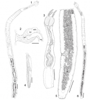
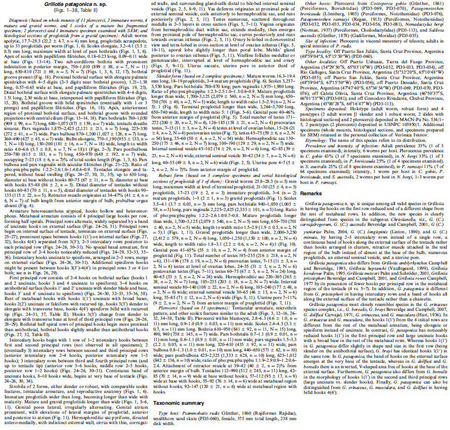
FIGURES 16. Grillotia patagonica n. sp. (1) Entire worm (robust form) ex Psammobatis rudis, scale bar = 1 mm. (2) Detail of terminal genitalia (slender form) ex P. rudis, scale bar=100 µm. (3) Pleroc... MoreFIGURES 16. Grillotia patagonica n. sp. (1) Entire worm (robust form) ex Psammobatis rudis, scale bar = 1 mm. (2) Detail of terminal genitalia (slender form) ex P. rudis, scale bar=100 µm. (3) Plerocercus ex Salilota australis, scale bar=1 mm. (4) Detail of the bulb from immature specimen ex P. rudis, scale bar=200 µm. (5) Terminal mature proglottid (slender form), dorsal view, scale bar=500 µm. (6) Entire worm (slender form) ex P. rudis, scale
bar = 1 mm. Circummedullar vitelline follicles not drawn in Figures 1 and 6, and partially drawn in Figure 5 to allow the view of internal organs. Abbreviations: esv, external seminal vesicle; hd, hermaphroditic duct; isv, internal seminal vesicle; vg, vagina. |
Line Drawing 2
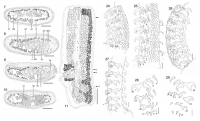
FIGURES 711. Grillotia patagonica n. sp. (710) Cross sections of a gravid proglottid (robust form), scale bar=250 µm. (7) At level of uterine pore. (8) At level of hermaphroditic sac (external semin... MoreFIGURES 711. Grillotia patagonica n. sp. (710) Cross sections of a gravid proglottid (robust form), scale bar=250 µm. (7) At level of uterine pore. (8) At level of hermaphroditic sac (external seminal vesicle). (9) At level of hermaphroditic sac (internal seminal vesicle). (10) At level of ovarian isthmus. (11) Gravid proglottid (robust form), ventral view, circummedullar vitelline follicles partially drawn to allow the view of internal organs, scale bar=500µm. Arrows indicate the level of cross sections in Figures 710. Abbreviations: c, cirrus; dod, dorsal osmoregulatory duct; esv, external seminal vesicle; hd, hermaphroditic duct; hs, hermaphroditic sac; isv, internal seminal vesicle; lm, longitudinal musculature; ov, ovary; t, testis; u, uterus; up, uterine pore; vg, vagina; vd, vas deferens; vf, vitelline follicles; vod, ventral osmoregulatory duct. FIGURES 2429. Tentacular armature of Grillotia patagonica n. sp. (24) Antibothrial surface (plerocercus ex Patagonotothen brevicauda), scale bar= 30 µm. (25) External surface (immature worm ex Psammobatis rudis), scale bar=30 µm. (26) Bothrial surface (plerocercus ex P. brevicauda), scale bar= 30 µm. (27) Internal surface (plerocercus ex P. brevicauda), scale bar=30 µm. (28) Profile of hooks along the tentacle, antibothrial surface (plerocercus ex Nemadactylus bergi), scale bar = 30 µm. (29) Profile of hooks along the tentacle, bothrial surface (plerocercus ex N. bergi), scale bar = 30 µm. Abbreviations: r1, row 1; r2, row 2; r3, row 3; r5, row 5. |
Photo Micrograph
|
Scanning Electron Micrograph
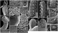
FIGURES 1223. Grilllotia patagonica n. sp., scanning electron micrographs. (12) Scolex, plerocercus ex Patagonotothen ramsayi, scale bar=200 µm; small numbers indicate locations of details shown in F... MoreFIGURES 1223. Grilllotia patagonica n. sp., scanning electron micrographs. (12) Scolex, plerocercus ex Patagonotothen ramsayi, scale bar=200 µm; small numbers indicate locations of details shown in Figures 13, 15, and 20. (13) Apex of scolex, scale bar=5 µm. (14) Detail of rounded projections with central cilium on the apex of the scolex, scale bar = 1 µm. (15) Anterior margin of the distal bothrial surface, scale bar = 2 µm. (16) Detail of bothrial groove, scale bar=5 µm. (17) Scolex of immature worm ex Psammobatis rudis, scale bar=200 µm; small numbers indicate locations of details shown in Figures 21 and 22. (18) Bothrial groove, anterior margin of bothrium, scale bar=500 µm; small numbers indicate locations of details shown in Figures 16 and 19. (19) Most-anterior proximal bothrial surface, scale bar = 2 µm. (20) Center of distal bothrial surface, scale bar = 2 µm. (21) Surface of pars vaginalis, scale bar=1 µm. (22) Surface of pars bulbosa, scale bar= 1 µm. (23) Bifid and trifid spinitriches covering the proximal bothrial surface near bothrial groove, scale bar = 1 µm. FIGURES 3038. Tentacular armature of Grillotia patagonica n. sp., scanning electron micrographs. (30) Antibothrial surface (plerocercus ex Patagonotothen ramsayi), scale bar=50 µm. (31) Bothrial surface (plerocercus ex P. ramsayi), scale bar=50 µm. (32) Internal surface (plerocercus ex P. ramsayi), scale bar = 50 µm. (33) First and second principal rows, bothrial surface (immature worm ex Psammobatis rudis), scale bar = 20 µm. (34) External surface (plerocercus ex Cottoperca gobio), scale bar = 30 µm. (35) Antibothrial surface (plerocercus ex C. gobio), scale bar = 50 µm. (36)
Antibothrial surface, detail of reduced hooks on first principal row (1040) (plerocercus ex P. ramsayi), scale bar = 10 µm. (37) Detail of bifid hook (4)
(plerocercus ex P. ramsayi), scale bar=4 µm. (38) Detail of reduced hook 10 on first row (antibothrial surface) (plerocercus ex P. ramsayi), scale bar=4
µm. |
 FIGURES 16. Grillotia patagonica n. sp. (1) Entire worm (robust form) ex Psammobatis rudis, scale bar = 1 mm. (2) Detail of terminal genitalia (slender form) ex P. rudis, scale bar=100 µm. (3) Plerocercus ex Salilota australis, scale bar=1 mm. (4) Detail of the bulb from immature specimen ex P. rudis, scale bar=200 µm. (5) Terminal mature proglottid (slender form), dorsal view, scale bar=500 µm. (6) Entire worm (slender form) ex P. rudis, scale
bar = 1 mm. Circummedullar vitelline follicles not drawn in Figures 1 and 6, and partially drawn in Figure 5 to allow the view of internal organs. Abbreviations: esv, external seminal vesicle; hd, hermaphroditic duct; isv, internal seminal vesicle; vg, vagina.
FIGURES 16. Grillotia patagonica n. sp. (1) Entire worm (robust form) ex Psammobatis rudis, scale bar = 1 mm. (2) Detail of terminal genitalia (slender form) ex P. rudis, scale bar=100 µm. (3) Plerocercus ex Salilota australis, scale bar=1 mm. (4) Detail of the bulb from immature specimen ex P. rudis, scale bar=200 µm. (5) Terminal mature proglottid (slender form), dorsal view, scale bar=500 µm. (6) Entire worm (slender form) ex P. rudis, scale
bar = 1 mm. Circummedullar vitelline follicles not drawn in Figures 1 and 6, and partially drawn in Figure 5 to allow the view of internal organs. Abbreviations: esv, external seminal vesicle; hd, hermaphroditic duct; isv, internal seminal vesicle; vg, vagina. 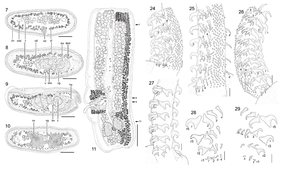 FIGURES 711. Grillotia patagonica n. sp. (710) Cross sections of a gravid proglottid (robust form), scale bar=250 µm. (7) At level of uterine pore. (8) At level of hermaphroditic sac (external seminal vesicle). (9) At level of hermaphroditic sac (internal seminal vesicle). (10) At level of ovarian isthmus. (11) Gravid proglottid (robust form), ventral view, circummedullar vitelline follicles partially drawn to allow the view of internal organs, scale bar=500µm. Arrows indicate the level of cross sections in Figures 710. Abbreviations: c, cirrus; dod, dorsal osmoregulatory duct; esv, external seminal vesicle; hd, hermaphroditic duct; hs, hermaphroditic sac; isv, internal seminal vesicle; lm, longitudinal musculature; ov, ovary; t, testis; u, uterus; up, uterine pore; vg, vagina; vd, vas deferens; vf, vitelline follicles; vod, ventral osmoregulatory duct. FIGURES 2429. Tentacular armature of Grillotia patagonica n. sp. (24) Antibothrial surface (plerocercus ex Patagonotothen brevicauda), scale bar= 30 µm. (25) External surface (immature worm ex Psammobatis rudis), scale bar=30 µm. (26) Bothrial surface (plerocercus ex P. brevicauda), scale bar= 30 µm. (27) Internal surface (plerocercus ex P. brevicauda), scale bar=30 µm. (28) Profile of hooks along the tentacle, antibothrial surface (plerocercus ex Nemadactylus bergi), scale bar = 30 µm. (29) Profile of hooks along the tentacle, bothrial surface (plerocercus ex N. bergi), scale bar = 30 µm. Abbreviations: r1, row 1; r2, row 2; r3, row 3; r5, row 5.
 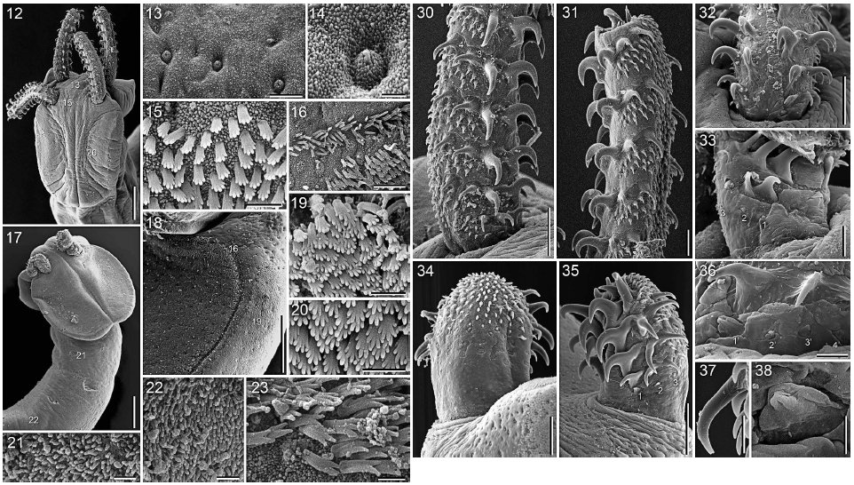 FIGURES 1223. Grilllotia patagonica n. sp., scanning electron micrographs. (12) Scolex, plerocercus ex Patagonotothen ramsayi, scale bar=200 µm; small numbers indicate locations of details shown in Figures 13, 15, and 20. (13) Apex of scolex, scale bar=5 µm. (14) Detail of rounded projections with central cilium on the apex of the scolex, scale bar = 1 µm. (15) Anterior margin of the distal bothrial surface, scale bar = 2 µm. (16) Detail of bothrial groove, scale bar=5 µm. (17) Scolex of immature worm ex Psammobatis rudis, scale bar=200 µm; small numbers indicate locations of details shown in Figures 21 and 22. (18) Bothrial groove, anterior margin of bothrium, scale bar=500 µm; small numbers indicate locations of details shown in Figures 16 and 19. (19) Most-anterior proximal bothrial surface, scale bar = 2 µm. (20) Center of distal bothrial surface, scale bar = 2 µm. (21) Surface of pars vaginalis, scale bar=1 µm. (22) Surface of pars bulbosa, scale bar= 1 µm. (23) Bifid and trifid spinitriches covering the proximal bothrial surface near bothrial groove, scale bar = 1 µm. FIGURES 3038. Tentacular armature of Grillotia patagonica n. sp., scanning electron micrographs. (30) Antibothrial surface (plerocercus ex Patagonotothen ramsayi), scale bar=50 µm. (31) Bothrial surface (plerocercus ex P. ramsayi), scale bar=50 µm. (32) Internal surface (plerocercus ex P. ramsayi), scale bar = 50 µm. (33) First and second principal rows, bothrial surface (immature worm ex Psammobatis rudis), scale bar = 20 µm. (34) External surface (plerocercus ex Cottoperca gobio), scale bar = 30 µm. (35) Antibothrial surface (plerocercus ex C. gobio), scale bar = 50 µm. (36)
Antibothrial surface, detail of reduced hooks on first principal row (1040) (plerocercus ex P. ramsayi), scale bar = 10 µm. (37) Detail of bifid hook (4)
(plerocercus ex P. ramsayi), scale bar=4 µm. (38) Detail of reduced hook 10 on first row (antibothrial surface) (plerocercus ex P. ramsayi), scale bar=4
µm.
FIGURES 1223. Grilllotia patagonica n. sp., scanning electron micrographs. (12) Scolex, plerocercus ex Patagonotothen ramsayi, scale bar=200 µm; small numbers indicate locations of details shown in Figures 13, 15, and 20. (13) Apex of scolex, scale bar=5 µm. (14) Detail of rounded projections with central cilium on the apex of the scolex, scale bar = 1 µm. (15) Anterior margin of the distal bothrial surface, scale bar = 2 µm. (16) Detail of bothrial groove, scale bar=5 µm. (17) Scolex of immature worm ex Psammobatis rudis, scale bar=200 µm; small numbers indicate locations of details shown in Figures 21 and 22. (18) Bothrial groove, anterior margin of bothrium, scale bar=500 µm; small numbers indicate locations of details shown in Figures 16 and 19. (19) Most-anterior proximal bothrial surface, scale bar = 2 µm. (20) Center of distal bothrial surface, scale bar = 2 µm. (21) Surface of pars vaginalis, scale bar=1 µm. (22) Surface of pars bulbosa, scale bar= 1 µm. (23) Bifid and trifid spinitriches covering the proximal bothrial surface near bothrial groove, scale bar = 1 µm. FIGURES 3038. Tentacular armature of Grillotia patagonica n. sp., scanning electron micrographs. (30) Antibothrial surface (plerocercus ex Patagonotothen ramsayi), scale bar=50 µm. (31) Bothrial surface (plerocercus ex P. ramsayi), scale bar=50 µm. (32) Internal surface (plerocercus ex P. ramsayi), scale bar = 50 µm. (33) First and second principal rows, bothrial surface (immature worm ex Psammobatis rudis), scale bar = 20 µm. (34) External surface (plerocercus ex Cottoperca gobio), scale bar = 30 µm. (35) Antibothrial surface (plerocercus ex C. gobio), scale bar = 50 µm. (36)
Antibothrial surface, detail of reduced hooks on first principal row (1040) (plerocercus ex P. ramsayi), scale bar = 10 µm. (37) Detail of bifid hook (4)
(plerocercus ex P. ramsayi), scale bar=4 µm. (38) Detail of reduced hook 10 on first row (antibothrial surface) (plerocercus ex P. ramsayi), scale bar=4
µm. 

