Line Drawing 1

Figure 7. Line drawings of Rhinebothrium brooksi n. sp. A. Scolex of Holotype (MZUSP 6124). B. Terminal mature proglottid of Paratype (USNPC
104712). Arrows indicate locations of sections shown in Fi... MoreFigure 7. Line drawings of Rhinebothrium brooksi n. sp. A. Scolex of Holotype (MZUSP 6124). B. Terminal mature proglottid of Paratype (USNPC
104712). Arrows indicate locations of sections shown in Figure. 10. C. Anterior and posterior portions of whole worm (Paratype, MZUSP 6123). Arrow
indicates anterior most mature proglottid. Abbreviations: CS, Cirrus sac; MG, Mehlis gland; O, Ovary; T, Testes; U, Uterus; V, Vitellaria; VA Vagina; VD,
vas deferens; VS Vaginal sphincter.
doi:10.1371/journal.pone.0022604.g007 |
Line Drawing 2

Figure. 2. Best-scoring ML tree (lnL = 7193.942769) based on COI data from marine and freshwater rhinebothriids. Numbers on internal branches denote nodal support as inferred by Bootstrap Proportions... MoreFigure. 2. Best-scoring ML tree (lnL = 7193.942769) based on COI data from marine and freshwater rhinebothriids. Numbers on internal branches denote nodal support as inferred by Bootstrap Proportions based on 5,000 replicates. *, indicates nodes recovered during phylogenetic analysis under parsimony. Numbers between square brackets in front of terminals represent total length in millimeters for those specimens measured. Scale indicates expected number of substitution per site. |
Photo Micrograph
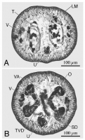
Figure 8. Cross-section through mature proglottid of Rhinebothrium
brooksi n. sp. A. Section at level of testes. B. Section at level of
ovarian isthmus. Abbreviations: LM, Longitudinal muscles; O, O... MoreFigure 8. Cross-section through mature proglottid of Rhinebothrium
brooksi n. sp. A. Section at level of testes. B. Section at level of
ovarian isthmus. Abbreviations: LM, Longitudinal muscles; O, Ovary; SD
Sperm duct; T, Testes; TVD, Transverse vitelline duct; U, Uterus; V,
Vitellaria; VA Vagina.
doi:10.1371/journal.pone.0022604.g008 |
Scanning Electron Micrograph
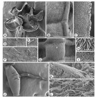
Figure 9. Scanning electron micrographs of Rhinebothrium brooksi n. sp. Scolex, Figures AI. Small letters indicate locations of details shown
in B and G. A. Scolex. B. Proximal surface of rim of bot... MoreFigure 9. Scanning electron micrographs of Rhinebothrium brooksi n. sp. Scolex, Figures AI. Small letters indicate locations of details shown
in B and G. A. Scolex. B. Proximal surface of rim of bothridium. Small letter indicates location of C. C. Proximal bothridial surface near bothridial rim. D.
Proximal surface near anterior of bothridium. E. Cilium on proximal bothridial surface. F. Proximal surface near middle of bothridium. G. Transverse
septum on distal bothridial surface. Small letter indicates location of H. H. Longitudinal septum. I. Stalk surface. Cirrus, Figures JL. J. Free proglottid
with everted cirrus. Small letters indicate location of K and L. K. Coniform spinithrix and capilliform filitriches on cirrus base. L. Coniform spinitriches
and capilliform filitriches on distal portion of cirrus. Scale bars: A, 200 mm; B, 20 mm; CF, 2 mm; G, 20 mm; HI, 2 mm; J, 200 mm; KL, 2 mm.
doi:10.1371/journal.pone.0022604.g009 |
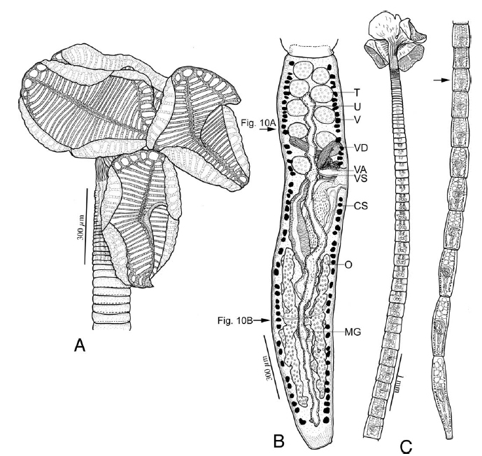 Figure 7. Line drawings of Rhinebothrium brooksi n. sp. A. Scolex of Holotype (MZUSP 6124). B. Terminal mature proglottid of Paratype (USNPC
104712). Arrows indicate locations of sections shown in Figure. 10. C. Anterior and posterior portions of whole worm (Paratype, MZUSP 6123). Arrow
indicates anterior most mature proglottid. Abbreviations: CS, Cirrus sac; MG, Mehlis gland; O, Ovary; T, Testes; U, Uterus; V, Vitellaria; VA Vagina; VD,
vas deferens; VS Vaginal sphincter.
doi:10.1371/journal.pone.0022604.g007
Figure 7. Line drawings of Rhinebothrium brooksi n. sp. A. Scolex of Holotype (MZUSP 6124). B. Terminal mature proglottid of Paratype (USNPC
104712). Arrows indicate locations of sections shown in Figure. 10. C. Anterior and posterior portions of whole worm (Paratype, MZUSP 6123). Arrow
indicates anterior most mature proglottid. Abbreviations: CS, Cirrus sac; MG, Mehlis gland; O, Ovary; T, Testes; U, Uterus; V, Vitellaria; VA Vagina; VD,
vas deferens; VS Vaginal sphincter.
doi:10.1371/journal.pone.0022604.g007 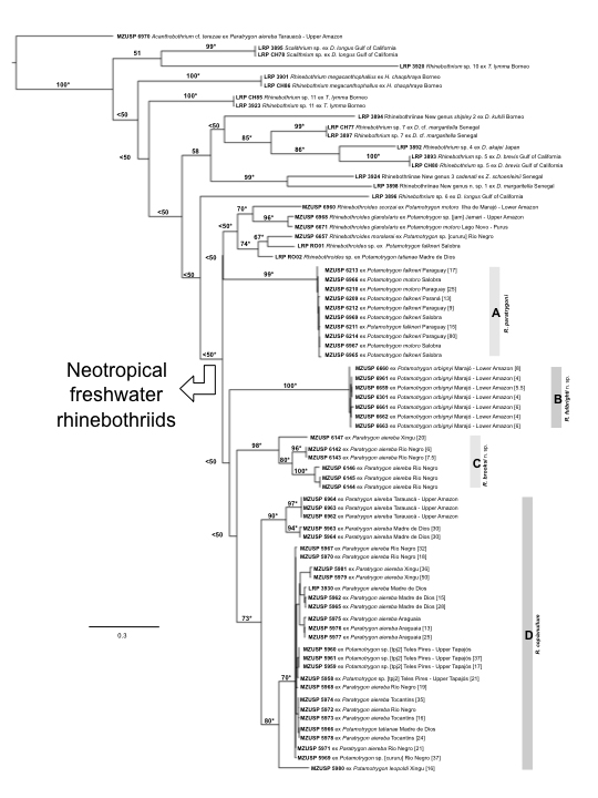 Figure. 2. Best-scoring ML tree (lnL = 7193.942769) based on COI data from marine and freshwater rhinebothriids. Numbers on internal branches denote nodal support as inferred by Bootstrap Proportions based on 5,000 replicates. *, indicates nodes recovered during phylogenetic analysis under parsimony. Numbers between square brackets in front of terminals represent total length in millimeters for those specimens measured. Scale indicates expected number of substitution per site.
 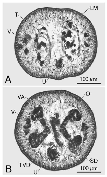 Figure 8. Cross-section through mature proglottid of Rhinebothrium
brooksi n. sp. A. Section at level of testes. B. Section at level of
ovarian isthmus. Abbreviations: LM, Longitudinal muscles; O, Ovary; SD
Sperm duct; T, Testes; TVD, Transverse vitelline duct; U, Uterus; V,
Vitellaria; VA Vagina.
doi:10.1371/journal.pone.0022604.g008
Figure 8. Cross-section through mature proglottid of Rhinebothrium
brooksi n. sp. A. Section at level of testes. B. Section at level of
ovarian isthmus. Abbreviations: LM, Longitudinal muscles; O, Ovary; SD
Sperm duct; T, Testes; TVD, Transverse vitelline duct; U, Uterus; V,
Vitellaria; VA Vagina.
doi:10.1371/journal.pone.0022604.g008  Figure 9. Scanning electron micrographs of Rhinebothrium brooksi n. sp. Scolex, Figures AI. Small letters indicate locations of details shown
in B and G. A. Scolex. B. Proximal surface of rim of bothridium. Small letter indicates location of C. C. Proximal bothridial surface near bothridial rim. D.
Proximal surface near anterior of bothridium. E. Cilium on proximal bothridial surface. F. Proximal surface near middle of bothridium. G. Transverse
septum on distal bothridial surface. Small letter indicates location of H. H. Longitudinal septum. I. Stalk surface. Cirrus, Figures JL. J. Free proglottid
with everted cirrus. Small letters indicate location of K and L. K. Coniform spinithrix and capilliform filitriches on cirrus base. L. Coniform spinitriches
and capilliform filitriches on distal portion of cirrus. Scale bars: A, 200 mm; B, 20 mm; CF, 2 mm; G, 20 mm; HI, 2 mm; J, 200 mm; KL, 2 mm.
doi:10.1371/journal.pone.0022604.g009
Figure 9. Scanning electron micrographs of Rhinebothrium brooksi n. sp. Scolex, Figures AI. Small letters indicate locations of details shown
in B and G. A. Scolex. B. Proximal surface of rim of bothridium. Small letter indicates location of C. C. Proximal bothridial surface near bothridial rim. D.
Proximal surface near anterior of bothridium. E. Cilium on proximal bothridial surface. F. Proximal surface near middle of bothridium. G. Transverse
septum on distal bothridial surface. Small letter indicates location of H. H. Longitudinal septum. I. Stalk surface. Cirrus, Figures JL. J. Free proglottid
with everted cirrus. Small letters indicate location of K and L. K. Coniform spinithrix and capilliform filitriches on cirrus base. L. Coniform spinitriches
and capilliform filitriches on distal portion of cirrus. Scale bars: A, 200 mm; B, 20 mm; CF, 2 mm; G, 20 mm; HI, 2 mm; J, 200 mm; KL, 2 mm.
doi:10.1371/journal.pone.0022604.g009 

