Line Drawing 1

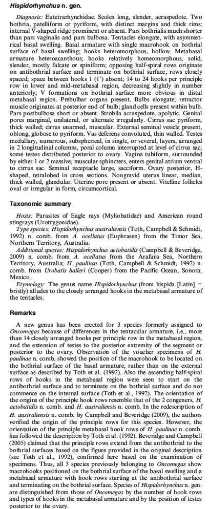
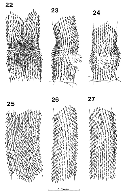
Figs. 22-27. Oncomegas paulinae n. sp., tentacular armature. 22. Basal armature and lower metabasal region of tentacle, internal face. 23. Basal region of tentacle, bothridial face, note asymmetrical ... MoreFigs. 22-27. Oncomegas paulinae n. sp., tentacular armature. 22. Basal armature and lower metabasal region of tentacle, internal face. 23. Basal region of tentacle, bothridial face, note asymmetrical swelling on internal face at left. 24. Basal region of tentacle, external face, note macrohook of basal armature. 25. Metabasal region, internal face. 26. Metabasal region, bothridial face. 27. Metabasal region, external face. |
Line Drawing 2

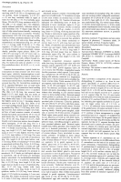
Figs. 28-32. Oncomegas paulinae n. sp. 28. Scolex. 29. Hook types from one file, proximal to distal, along internal face: (a-e) basal armature, (f-h) lower metabasal to mid-region of tentacle. 30. Hoo... MoreFigs. 28-32. Oncomegas paulinae n. sp. 28. Scolex. 29. Hook types from one file, proximal to distal, along internal face: (a-e) basal armature, (f-h) lower metabasal to mid-region of tentacle. 30. Hooks types from one file, proximal to distal, along bothridial face: (a-c) basal armature, (d-f) lower metabasal to mid-region of tentacle. 31. Hook types from one file, proximal to distal on external face: (a-e) basal armature, (f-i) lower metabasal to mid-region of tentacle. 32. Anterior portion of bulb showing prebulbar organ. Figs. 33-39. Oncomegas paulinae n. sp. 33. Mature segment. 34. Gravid segment. Figs. 35-37. Transverse sections through mature segment. 35. Proximal to cirrus-sac; note dorsal interruption of vitelline follicles. 36. Through terminal genitalia; note vaginal sphincters and position of osmoregulatory canals dorsal to cirrus-sac. 37. Section through ovary at ovarian isthmus; note lateral position of osmoregulatory ducts. 38. Embryonated egg, showing conical projection attached to oncospheral membrane. 39. Dorsal view of cirrus-sac, external seminal vesicle and distal vagina. Abbreviations: C, conical projection; CS, cirrus-sac; DC, dorsal osmoregulatory canal; DVS, distal vaginal sphincter; ESV, external seminal vesicle; LM, longitudinal muscle bundles; OV, ovary; PVS, proximal vaginal sphincter; SD, sperm duct; T, testis; U, uterus; V, vagina; VC, ventral osmoregulatory canal; VD, vas deferens; VT, vitelline follicle. |
Photo Micrograph
|
Scanning Electron Micrograph
|
 Figs. 22-27. Oncomegas paulinae n. sp., tentacular armature. 22. Basal armature and lower metabasal region of tentacle, internal face. 23. Basal region of tentacle, bothridial face, note asymmetrical swelling on internal face at left. 24. Basal region of tentacle, external face, note macrohook of basal armature. 25. Metabasal region, internal face. 26. Metabasal region, bothridial face. 27. Metabasal region, external face.
 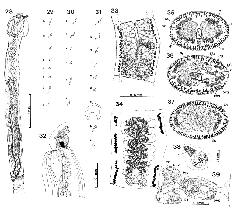 Figs. 28-32. Oncomegas paulinae n. sp. 28. Scolex. 29. Hook types from one file, proximal to distal, along internal face: (a-e) basal armature, (f-h) lower metabasal to mid-region of tentacle. 30. Hooks types from one file, proximal to distal, along bothridial face: (a-c) basal armature, (d-f) lower metabasal to mid-region of tentacle. 31. Hook types from one file, proximal to distal on external face: (a-e) basal armature, (f-i) lower metabasal to mid-region of tentacle. 32. Anterior portion of bulb showing prebulbar organ. Figs. 33-39. Oncomegas paulinae n. sp. 33. Mature segment. 34. Gravid segment. Figs. 35-37. Transverse sections through mature segment. 35. Proximal to cirrus-sac; note dorsal interruption of vitelline follicles. 36. Through terminal genitalia; note vaginal sphincters and position of osmoregulatory canals dorsal to cirrus-sac. 37. Section through ovary at ovarian isthmus; note lateral position of osmoregulatory ducts. 38. Embryonated egg, showing conical projection attached to oncospheral membrane. 39. Dorsal view of cirrus-sac, external seminal vesicle and distal vagina. Abbreviations: C, conical projection; CS, cirrus-sac; DC, dorsal osmoregulatory canal; DVS, distal vaginal sphincter; ESV, external seminal vesicle; LM, longitudinal muscle bundles; OV, ovary; PVS, proximal vaginal sphincter; SD, sperm duct; T, testis; U, uterus; V, vagina; VC, ventral osmoregulatory canal; VD, vas deferens; VT, vitelline follicle.