Line Drawing 1
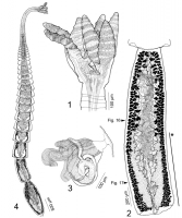
FIGURES 14. Line drawings of Crassuseptum pietrafacei n. gen., n. sp. (1) Scolex (Holotype). (2) Mature proglottid (Paratype); note that for clarity, only 1 of 23 layers of testes throughout the pro... MoreFIGURES 14. Line drawings of Crassuseptum pietrafacei n. gen., n. sp. (1) Scolex (Holotype). (2) Mature proglottid (Paratype); note that for clarity, only 1 of 23 layers of testes throughout the proglottid are drawn. Arrows indicate location of cross sections shown in Figures 1617. Asterisk (*) indicates portion of proglottid, corresponding to ovary, in which not all testes are drawn. (3) Detail of terminal genitalia (Paratype). (4) Whole
worm (Paratype). |
Line Drawing 2
|
Photo Micrograph

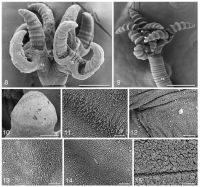
FIGURES 57. Histological and optical sections of scolex of Crassuseptum pietrafacei n. gen., n. sp. (5) Optical, longitudinal section through anterior
portion of bothridium (USNPC No. 105266). (6) H... MoreFIGURES 57. Histological and optical sections of scolex of Crassuseptum pietrafacei n. gen., n. sp. (5) Optical, longitudinal section through anterior
portion of bothridium (USNPC No. 105266). (6) Histological, longitudinal section through anterior portion of bothridium (LRP No. 7796). (7) Histological, longitudinal section through middle of bothridium (LRP No. 7795). Abbreviations: m, middle region of bothridium; r, radial musculature;
s, septal musculature. Asterisk (*) denotes 2 reduced septa on each side of middle of bothridium. Scale bars: 50 µm. FIGURES 1617. Histological sections of proglottid of Crassuseptum pietrafacei n. gen., n. sp. (16) Cross section at level of testes. (17) Cross section at level of ovarian isthmus (LRP No. 7787). Abbreviations: DED, dorsal excretory duct; O, ovary; T, testis; U, uterus; VA, vagina; VD, vas deferens; VED,
ventral excretory duct; VI, vitellaria. Scale bars: 100 µm. |
Scanning Electron Micrograph
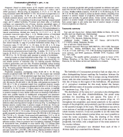
FIGURES 815. Scanning electron micrographs (SEMs) of Crassuseptum pietrafacei n. gen., n. sp. (8) Scolex. Small numbers indicate locations of detail shown in Figures 1114. (9) Scolex. Small numbers ... MoreFIGURES 815. Scanning electron micrographs (SEMs) of Crassuseptum pietrafacei n. gen., n. sp. (8) Scolex. Small numbers indicate locations of detail shown in Figures 1114. (9) Scolex. Small numbers indicate locations of detail shown in Figs. 10, 15. (10) Anterior loculus distal bothridial
surface. (11) Other loculus on distal bothridial surface. (12) Middle, nonloculated, portion of bothridium. (13) Proximal bothridial surface. (14) Stalk,
note cilium. (15) Strobila. Scale bars: 8, 100 µm; 9, 200 µm; 10, 10 µm; 1115, 2 µm. |
 FIGURES 14. Line drawings of Crassuseptum pietrafacei n. gen., n. sp. (1) Scolex (Holotype). (2) Mature proglottid (Paratype); note that for clarity, only 1 of 23 layers of testes throughout the proglottid are drawn. Arrows indicate location of cross sections shown in Figures 1617. Asterisk (*) indicates portion of proglottid, corresponding to ovary, in which not all testes are drawn. (3) Detail of terminal genitalia (Paratype). (4) Whole
worm (Paratype).
FIGURES 14. Line drawings of Crassuseptum pietrafacei n. gen., n. sp. (1) Scolex (Holotype). (2) Mature proglottid (Paratype); note that for clarity, only 1 of 23 layers of testes throughout the proglottid are drawn. Arrows indicate location of cross sections shown in Figures 1617. Asterisk (*) indicates portion of proglottid, corresponding to ovary, in which not all testes are drawn. (3) Detail of terminal genitalia (Paratype). (4) Whole
worm (Paratype). 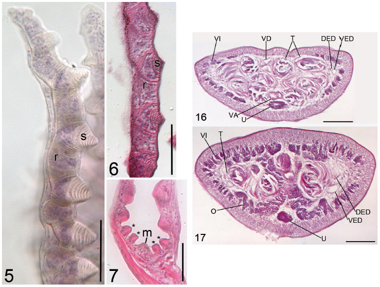 FIGURES 57. Histological and optical sections of scolex of Crassuseptum pietrafacei n. gen., n. sp. (5) Optical, longitudinal section through anterior
portion of bothridium (USNPC No. 105266). (6) Histological, longitudinal section through anterior portion of bothridium (LRP No. 7796). (7) Histological, longitudinal section through middle of bothridium (LRP No. 7795). Abbreviations: m, middle region of bothridium; r, radial musculature;
s, septal musculature. Asterisk (*) denotes 2 reduced septa on each side of middle of bothridium. Scale bars: 50 µm. FIGURES 1617. Histological sections of proglottid of Crassuseptum pietrafacei n. gen., n. sp. (16) Cross section at level of testes. (17) Cross section at level of ovarian isthmus (LRP No. 7787). Abbreviations: DED, dorsal excretory duct; O, ovary; T, testis; U, uterus; VA, vagina; VD, vas deferens; VED,
ventral excretory duct; VI, vitellaria. Scale bars: 100 µm.
FIGURES 57. Histological and optical sections of scolex of Crassuseptum pietrafacei n. gen., n. sp. (5) Optical, longitudinal section through anterior
portion of bothridium (USNPC No. 105266). (6) Histological, longitudinal section through anterior portion of bothridium (LRP No. 7796). (7) Histological, longitudinal section through middle of bothridium (LRP No. 7795). Abbreviations: m, middle region of bothridium; r, radial musculature;
s, septal musculature. Asterisk (*) denotes 2 reduced septa on each side of middle of bothridium. Scale bars: 50 µm. FIGURES 1617. Histological sections of proglottid of Crassuseptum pietrafacei n. gen., n. sp. (16) Cross section at level of testes. (17) Cross section at level of ovarian isthmus (LRP No. 7787). Abbreviations: DED, dorsal excretory duct; O, ovary; T, testis; U, uterus; VA, vagina; VD, vas deferens; VED,
ventral excretory duct; VI, vitellaria. Scale bars: 100 µm.  FIGURES 815. Scanning electron micrographs (SEMs) of Crassuseptum pietrafacei n. gen., n. sp. (8) Scolex. Small numbers indicate locations of detail shown in Figures 1114. (9) Scolex. Small numbers indicate locations of detail shown in Figs. 10, 15. (10) Anterior loculus distal bothridial
surface. (11) Other loculus on distal bothridial surface. (12) Middle, nonloculated, portion of bothridium. (13) Proximal bothridial surface. (14) Stalk,
note cilium. (15) Strobila. Scale bars: 8, 100 µm; 9, 200 µm; 10, 10 µm; 1115, 2 µm.
FIGURES 815. Scanning electron micrographs (SEMs) of Crassuseptum pietrafacei n. gen., n. sp. (8) Scolex. Small numbers indicate locations of detail shown in Figures 1114. (9) Scolex. Small numbers indicate locations of detail shown in Figs. 10, 15. (10) Anterior loculus distal bothridial
surface. (11) Other loculus on distal bothridial surface. (12) Middle, nonloculated, portion of bothridium. (13) Proximal bothridial surface. (14) Stalk,
note cilium. (15) Strobila. Scale bars: 8, 100 µm; 9, 200 µm; 10, 10 µm; 1115, 2 µm. 

