Line Drawing 1
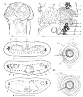
Figs. 510. Ophiotaenia georgievi sp. n. Fig. 5. Holotype, scolex, lateral view (MHNG INVE 65470). Fig. 6. Paratype, vagina and
cirrus-sac region, dorsal view (MHNG INVE 65473). Figs. 79. Cross-sect... MoreFigs. 510. Ophiotaenia georgievi sp. n. Fig. 5. Holotype, scolex, lateral view (MHNG INVE 65470). Fig. 6. Paratype, vagina and
cirrus-sac region, dorsal view (MHNG INVE 65473). Figs. 79. Cross-sections at the level of the ovary, the anterior part and the
posterior part of the testicular region, respectively (MHNG INVE 65475). Fig. 10. Eggs, drawn in distilled water, showing the three-layered embryophore (MHNG INVE 65475); an additional layer marked by an arrow. Abbreviations: cg - cells with finely granular cytoplasm; ci cirrus; cs cirrus-sac; do dorsal osmoregulatory canal; du uterine diverticles; em embryophore; lm internal longitudinal musculature; oe outer envelope; on oncosphere; ov ovary; sc secondary canals; te testes; ut uterus; vc vaginal canal; vd vas deferens; vi vitelline follicles; vo ventral osmoregulatory canals; vs vaginal sphincter. Scale bars:
Fig. 5 = 100 μm; Figs. 69 = 250 μm; Fig. 10 = 50 μm. |
Line Drawing 2
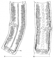
Figs. 11, 12. Ophiotaenia georgievi sp. n. Fig. 11. Holotype, mature proglottis, dorsal view (MHNG INVE 65470). Fig. 12. Paratype,
pregravid proglottis, dorsal view (MHNG INVE 65473). Abbreviations: ... MoreFigs. 11, 12. Ophiotaenia georgievi sp. n. Fig. 11. Holotype, mature proglottis, dorsal view (MHNG INVE 65470). Fig. 12. Paratype,
pregravid proglottis, dorsal view (MHNG INVE 65473). Abbreviations: mg Mehlis glands. Scale bars = 500 μm. |
Photo Micrograph

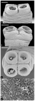
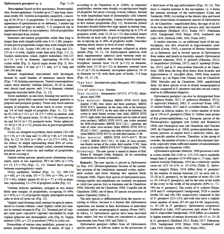
Figs. 13, 14. Ophiotaenia georgievi sp. n. Eggs in distilled water, showing the three-layered
embryophore (MHNG INVE 65475); an additional layer marked by an arrow. Abbreviations: em = embryopho... MoreFigs. 13, 14. Ophiotaenia georgievi sp. n. Eggs in distilled water, showing the three-layered
embryophore (MHNG INVE 65475); an additional layer marked by an arrow. Abbreviations: em = embryophore; oe = outer envelope; on = oncosphere. Scale bars = 20 μm. |
Scanning Electron Micrograph
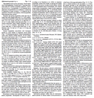
Figs. 14. Ophiotaenia georgievi sp. n.; scanning electron micrographs. Paratype (MHNG INVE 65474). Fig. 1. Scolex, dorsoventral view. Fig. 2. Scolex, lateral view. Fig. 3. Scolex, apical
view. ... MoreFigs. 14. Ophiotaenia georgievi sp. n.; scanning electron micrographs. Paratype (MHNG INVE 65474). Fig. 1. Scolex, dorsoventral view. Fig. 2. Scolex, lateral view. Fig. 3. Scolex, apical
view. Fig. 4. Microtriches at the level of the apex of scolex. Scale bars: Figs. 13 = 50 μm; Fig. 4 = 3 μm. |

 Figs. 510. Ophiotaenia georgievi sp. n. Fig. 5. Holotype, scolex, lateral view (MHNG INVE 65470). Fig. 6. Paratype, vagina and
cirrus-sac region, dorsal view (MHNG INVE 65473). Figs. 79. Cross-sections at the level of the ovary, the anterior part and the
posterior part of the testicular region, respectively (MHNG INVE 65475). Fig. 10. Eggs, drawn in distilled water, showing the three-layered embryophore (MHNG INVE 65475); an additional layer marked by an arrow. Abbreviations: cg - cells with finely granular cytoplasm; ci cirrus; cs cirrus-sac; do dorsal osmoregulatory canal; du uterine diverticles; em embryophore; lm internal longitudinal musculature; oe outer envelope; on oncosphere; ov ovary; sc secondary canals; te testes; ut uterus; vc vaginal canal; vd vas deferens; vi vitelline follicles; vo ventral osmoregulatory canals; vs vaginal sphincter. Scale bars:
Fig. 5 = 100 μm; Figs. 69 = 250 μm; Fig. 10 = 50 μm.
Figs. 510. Ophiotaenia georgievi sp. n. Fig. 5. Holotype, scolex, lateral view (MHNG INVE 65470). Fig. 6. Paratype, vagina and
cirrus-sac region, dorsal view (MHNG INVE 65473). Figs. 79. Cross-sections at the level of the ovary, the anterior part and the
posterior part of the testicular region, respectively (MHNG INVE 65475). Fig. 10. Eggs, drawn in distilled water, showing the three-layered embryophore (MHNG INVE 65475); an additional layer marked by an arrow. Abbreviations: cg - cells with finely granular cytoplasm; ci cirrus; cs cirrus-sac; do dorsal osmoregulatory canal; du uterine diverticles; em embryophore; lm internal longitudinal musculature; oe outer envelope; on oncosphere; ov ovary; sc secondary canals; te testes; ut uterus; vc vaginal canal; vd vas deferens; vi vitelline follicles; vo ventral osmoregulatory canals; vs vaginal sphincter. Scale bars:
Fig. 5 = 100 μm; Figs. 69 = 250 μm; Fig. 10 = 50 μm.  Figs. 11, 12. Ophiotaenia georgievi sp. n. Fig. 11. Holotype, mature proglottis, dorsal view (MHNG INVE 65470). Fig. 12. Paratype,
pregravid proglottis, dorsal view (MHNG INVE 65473). Abbreviations: mg Mehlis glands. Scale bars = 500 μm.
Figs. 11, 12. Ophiotaenia georgievi sp. n. Fig. 11. Holotype, mature proglottis, dorsal view (MHNG INVE 65470). Fig. 12. Paratype,
pregravid proglottis, dorsal view (MHNG INVE 65473). Abbreviations: mg Mehlis glands. Scale bars = 500 μm.  Figs. 13, 14. Ophiotaenia georgievi sp. n. Eggs in distilled water, showing the three-layered
embryophore (MHNG INVE 65475); an additional layer marked by an arrow. Abbreviations: em = embryophore; oe = outer envelope; on = oncosphere. Scale bars = 20 μm.
Figs. 13, 14. Ophiotaenia georgievi sp. n. Eggs in distilled water, showing the three-layered
embryophore (MHNG INVE 65475); an additional layer marked by an arrow. Abbreviations: em = embryophore; oe = outer envelope; on = oncosphere. Scale bars = 20 μm.  Figs. 14. Ophiotaenia georgievi sp. n.; scanning electron micrographs. Paratype (MHNG INVE 65474). Fig. 1. Scolex, dorsoventral view. Fig. 2. Scolex, lateral view. Fig. 3. Scolex, apical
view. Fig. 4. Microtriches at the level of the apex of scolex. Scale bars: Figs. 13 = 50 μm; Fig. 4 = 3 μm.
Figs. 14. Ophiotaenia georgievi sp. n.; scanning electron micrographs. Paratype (MHNG INVE 65474). Fig. 1. Scolex, dorsoventral view. Fig. 2. Scolex, lateral view. Fig. 3. Scolex, apical
view. Fig. 4. Microtriches at the level of the apex of scolex. Scale bars: Figs. 13 = 50 μm; Fig. 4 = 3 μm. 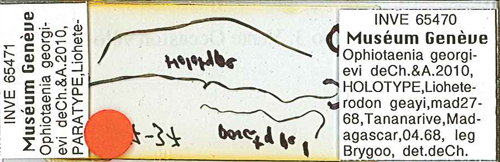 Holotype: MNHG-INVE No. 65470

