Line Drawing 1

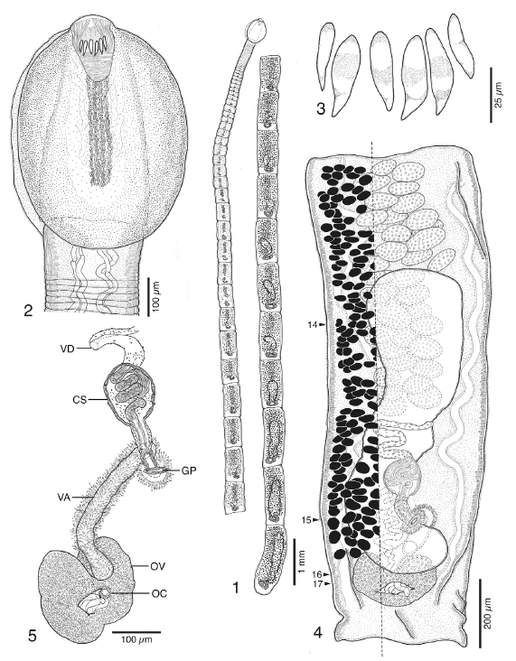
FIGURES 15. Line drawings of Ahamulina catarina n. gen., n. sp. from Scyliorhinus besnardi. 1. Holotype (MZUSP 6487a).
2. Scolex of holotype (MZUSP 6487a). 3. Detail of apical hooks of holotype (MZU... MoreFIGURES 15. Line drawings of Ahamulina catarina n. gen., n. sp. from Scyliorhinus besnardi. 1. Holotype (MZUSP 6487a).
2. Scolex of holotype (MZUSP 6487a). 3. Detail of apical hooks of holotype (MZUSP 6487a). 4. Gravid proglottid of paratype,
eggs not shown (USNPC 105708); small numbers at arrows indicate location at which sections in Figs. 1417 were taken. 5.
Detail of terminal genitalia of gravid proglottid of the paratype (USNPC 105708). Abbreviations: CS, cirrus sac; GP, genital
pore; OC, ovicapt; OV, ovary; VA, vagina; VD, vas deferens. |
Line Drawing 2
|
Photo Micrograph
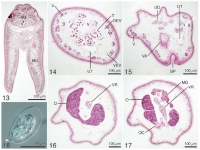
FIGURES 1318. Ahamulina catarina n. gen. n. sp. from Scyliorhinus besnardi. Figs 1317. Histological sections. 13. Sagittal
section through scolex showing bothria free posteriorly for most of their ... MoreFIGURES 1318. Ahamulina catarina n. gen. n. sp. from Scyliorhinus besnardi. Figs 1317. Histological sections. 13. Sagittal
section through scolex showing bothria free posteriorly for most of their length. 14. Cross section through proglottid at level of
testes. 15. Cross section through proglottid at level of genital pore. 16. Cross section through proglottid at level of ovarian
bridge. 17. Cross section through proglottid posterior to ovarian bridge. 18. Whole mount of egg; arrow indicates subterminal
knob. Abbreviations: AO, apical organ; BO, bothrium; DEV, dorsal excretory vessel; GP, genital pore; MG, Mehlis gland; O,
ovary; OC, ovicapt; T, testis; UD, uterine duct; UT, uterus; V, vitelline follicle; VA, vagina; VEV, ventral excretory vessel; VR,
vitelline reservoir. |
Scanning Electron Micrograph
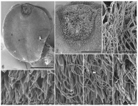
FIGURES 612. Scanning electron micrographs of Ahamulina catarina n. gen. n. sp. from Scyliorhinus besnardi. 6. Scolex;
small numbers indicate location of microthrix images in Figs. 912. 7. Detail o... MoreFIGURES 612. Scanning electron micrographs of Ahamulina catarina n. gen. n. sp. from Scyliorhinus besnardi. 6. Scolex;
small numbers indicate location of microthrix images in Figs. 912. 7. Detail of apical organ; small number indicates location
of detail in Fig. 8. 8. Enlarged view of surface of apical organ. 9. Enlarged view of trifurcate spinitriches on distal bothrial surface.
10. Enlarged view of trifurcate spinitriches on proximal bothrial surface (see arrow). 11. Enlarged view of cephalic peduncle.
12. Enlarged view of anterior of strobila. |
 FIGURES 15. Line drawings of Ahamulina catarina n. gen., n. sp. from Scyliorhinus besnardi. 1. Holotype (MZUSP 6487a).
2. Scolex of holotype (MZUSP 6487a). 3. Detail of apical hooks of holotype (MZUSP 6487a). 4. Gravid proglottid of paratype,
eggs not shown (USNPC 105708); small numbers at arrows indicate location at which sections in Figs. 1417 were taken. 5.
Detail of terminal genitalia of gravid proglottid of the paratype (USNPC 105708). Abbreviations: CS, cirrus sac; GP, genital
pore; OC, ovicapt; OV, ovary; VA, vagina; VD, vas deferens.
FIGURES 15. Line drawings of Ahamulina catarina n. gen., n. sp. from Scyliorhinus besnardi. 1. Holotype (MZUSP 6487a).
2. Scolex of holotype (MZUSP 6487a). 3. Detail of apical hooks of holotype (MZUSP 6487a). 4. Gravid proglottid of paratype,
eggs not shown (USNPC 105708); small numbers at arrows indicate location at which sections in Figs. 1417 were taken. 5.
Detail of terminal genitalia of gravid proglottid of the paratype (USNPC 105708). Abbreviations: CS, cirrus sac; GP, genital
pore; OC, ovicapt; OV, ovary; VA, vagina; VD, vas deferens. 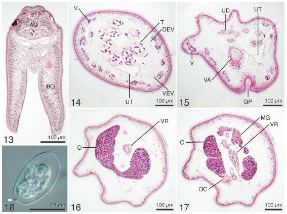 FIGURES 1318. Ahamulina catarina n. gen. n. sp. from Scyliorhinus besnardi. Figs 1317. Histological sections. 13. Sagittal
section through scolex showing bothria free posteriorly for most of their length. 14. Cross section through proglottid at level of
testes. 15. Cross section through proglottid at level of genital pore. 16. Cross section through proglottid at level of ovarian
bridge. 17. Cross section through proglottid posterior to ovarian bridge. 18. Whole mount of egg; arrow indicates subterminal
knob. Abbreviations: AO, apical organ; BO, bothrium; DEV, dorsal excretory vessel; GP, genital pore; MG, Mehlis gland; O,
ovary; OC, ovicapt; T, testis; UD, uterine duct; UT, uterus; V, vitelline follicle; VA, vagina; VEV, ventral excretory vessel; VR,
vitelline reservoir.
FIGURES 1318. Ahamulina catarina n. gen. n. sp. from Scyliorhinus besnardi. Figs 1317. Histological sections. 13. Sagittal
section through scolex showing bothria free posteriorly for most of their length. 14. Cross section through proglottid at level of
testes. 15. Cross section through proglottid at level of genital pore. 16. Cross section through proglottid at level of ovarian
bridge. 17. Cross section through proglottid posterior to ovarian bridge. 18. Whole mount of egg; arrow indicates subterminal
knob. Abbreviations: AO, apical organ; BO, bothrium; DEV, dorsal excretory vessel; GP, genital pore; MG, Mehlis gland; O,
ovary; OC, ovicapt; T, testis; UD, uterine duct; UT, uterus; V, vitelline follicle; VA, vagina; VEV, ventral excretory vessel; VR,
vitelline reservoir.  FIGURES 612. Scanning electron micrographs of Ahamulina catarina n. gen. n. sp. from Scyliorhinus besnardi. 6. Scolex;
small numbers indicate location of microthrix images in Figs. 912. 7. Detail of apical organ; small number indicates location
of detail in Fig. 8. 8. Enlarged view of surface of apical organ. 9. Enlarged view of trifurcate spinitriches on distal bothrial surface.
10. Enlarged view of trifurcate spinitriches on proximal bothrial surface (see arrow). 11. Enlarged view of cephalic peduncle.
12. Enlarged view of anterior of strobila.
FIGURES 612. Scanning electron micrographs of Ahamulina catarina n. gen. n. sp. from Scyliorhinus besnardi. 6. Scolex;
small numbers indicate location of microthrix images in Figs. 912. 7. Detail of apical organ; small number indicates location
of detail in Fig. 8. 8. Enlarged view of surface of apical organ. 9. Enlarged view of trifurcate spinitriches on distal bothrial surface.
10. Enlarged view of trifurcate spinitriches on proximal bothrial surface (see arrow). 11. Enlarged view of cephalic peduncle.
12. Enlarged view of anterior of strobila. 

