Cestode Scientific Name
| Species ID | 9652 |
|---|---|
| Order | Phyllobothriidea |
| Family | |
| Subfamily | |
| Genus | Trilocularia |
| Species | eberti |
| Authority | Pickering & Caira, 2012 |
| Taxonomic Status | Valid |
| Valid Name | |
| Synonyms | |
| Genus Record | No |
| Type Species | |
| Verified | No |
| Verified By | |
| Citation(s) |
Pickering, M. and J. N. Caira. 2012. A new hyperapolytic species, Trilocularia eberti sp. n. (Cestoda: Tetraphyllidea), from Squalus cf. mitsukurii (Squaliformes: Squalidae) off South Africa with comments on its development and fecundity. Folia Parasitologica 59(2): 107-114. (5839) Download PDF |
| Redescription | |
| Scientific Name Notes |
Record Data
| Date (MM/DD/YYYY) | Action | User Name |
|---|---|---|
| 06/20/2012 | Created | B. Barbeau |
| 01/02/2014 | Modified | |
| 06/06/2015 | Modified | J. Caira |
Type Host
| Host Class | |||||||
|---|---|---|---|---|---|---|---|
| Host Order | Squaliformes | ||||||
| Host Family | Squalidae | ||||||
|
Type Host (Literal) |
|
||||||
|
Type Host (Valid) |
|
||||||
| Additional Host(s) | |||||||
| Site in Host | |||||||
| Host Notes |
Type Locality
| Country | |
|---|---|
| Body of Water | |
| Island(s) | |
| City/Region | |
| Coordinates | |
| DD Latitude | |
| DD Longitude | |
| Additional Localities | |
| Locality Notes |
Specimens
| Type Material | |
|---|---|
| Total Number of Type Specimens | |
| Voucher Material | |
| Specimen Notes |
Data are given as in original description unless otherwise indicated.
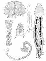
 Figs. 27. Line drawings of Trilocularia eberti sp. n. Fig. 2. Whole worm. Fig. 3. Scolex. Fig. 4. Dehisced free proglottid, ventral view. Arrowheads indicate location of cross-sections shown in Figs. 1722. Fig. 5. Anterior attachment region of free proglottid, dorsal view. Fig. 6. Detail of posterior portion of mature proglottid: cirrus sac and vagina, dorsal view. Fig. 7. Sketch of male reproductive system in free proglottid, dorsal view.
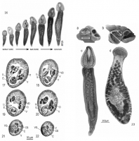
  Fig. 16. Light micrographs of free proglottids of Trilocularia eberti sp. n. representing the spectrum of developmental stages found. Figs. 1722. Histological sections of Trilocularia eberti sp. n. Fig. 17. Cross-section through testes. Fig. 18. Cross-section through testes and extensive vas deferens. Fig. 19. Cross-section through ovary arms (bilobed) and extensive vas deferens. Fig. 20. Cross-section through ovary. Fig. 21. Cross-section through cirrus sac and vagina. Fig. 22. Cross-section through cirrus sac and vagina close to genital pore opening. Abbreviations: C cirrus; CS cirrus sac; E eggs; O ovary; T testes; U uterus; V vitelline follicle; VD vas deferens; VG vagina. Fig. 23 ad. Trilocularia eberti sp. n. scolex (a) and free proglottid (c). Trilocularia gracilis scolex (b) and free proglottid (d) for comparison . White arrows on scoleces (a, b) illustrate difference in ration of anterior loculus to posterior loculi between the two species. a, b scanning electron micrographs (scoleces);
c, d light micrographs (free proglottids).
Fig. 16. Light micrographs of free proglottids of Trilocularia eberti sp. n. representing the spectrum of developmental stages found. Figs. 1722. Histological sections of Trilocularia eberti sp. n. Fig. 17. Cross-section through testes. Fig. 18. Cross-section through testes and extensive vas deferens. Fig. 19. Cross-section through ovary arms (bilobed) and extensive vas deferens. Fig. 20. Cross-section through ovary. Fig. 21. Cross-section through cirrus sac and vagina. Fig. 22. Cross-section through cirrus sac and vagina close to genital pore opening. Abbreviations: C cirrus; CS cirrus sac; E eggs; O ovary; T testes; U uterus; V vitelline follicle; VD vas deferens; VG vagina. Fig. 23 ad. Trilocularia eberti sp. n. scolex (a) and free proglottid (c). Trilocularia gracilis scolex (b) and free proglottid (d) for comparison . White arrows on scoleces (a, b) illustrate difference in ration of anterior loculus to posterior loculi between the two species. a, b scanning electron micrographs (scoleces);
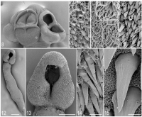
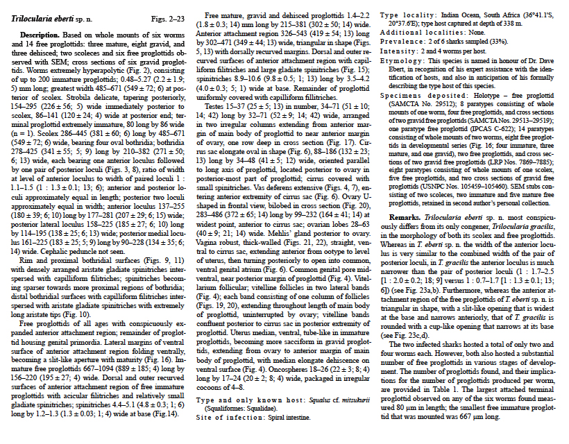
c, d light micrographs (free proglottids).  Figs. 815. Scanning electron micrographs of Trilocularia eberti sp. n. Note: small numbers in Figs. 8 and 13 correpsond to Figs. 9-11 and 15. Fig. 8. Scolex. Fig. 9. Detail of proximal surface of anterior loculus. Fig. 10. Detail of distal surface of medial posterior loculus. Inset: Enlarged view showing long aristate tips on gladiate spinitriches. Fig. 11. Detail of proximal surface of medial posterior loculus. Fig. 12. Mature free proglottid. Fig. 13. Detail of anterior attachment region of mature free proglottid. Fig. 14. Detail of outer surface of anterior attachment region of immature free proglottid. Fig. 15. Detail of outer surface of anterior attachment region of mature free proglottid. Scale bars: Figs. 8, 12 = 200 µm; Figs. 9-11, 14, 15 = 2 µm; Inset of Fig. 10 = 1 µm; Fig. 13 = 100 µm.
 Best viewed in Firefox


