Cestode Scientific Name
| Species ID | 9071 |
|---|---|
| Order | Cyclophyllidea |
| Family | Paruterinidae |
| Subfamily | |
| Genus | Anonchotaenia |
| Species | rostellata |
| Authority | Rodgers, 1941 |
| Taxonomic Status | |
| Valid Name | |
| Synonyms | Orthoskrjabinia rostellata (Rodgers, 1941) |
| Genus Record | No |
| Type Species | No |
| Verified | No |
| Verified By | |
| Citation(s) |
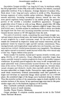
Rodgers, L. O. 1941. A new dilepidid tapeworm from a cardinal. Transactions of the American Microscopical Society 60(2): 273-275. (5484) Download PDF |
| Redescription | |
| Scientific Name Notes | Considered by Spassky (1977) as probable synonym of Orthoskrjabinia conica. A. costellata appears to be a misspelling of A. rostellata Rodgers, 1941. This misspelling appears in the Global Names Index (globalnames.org). |
Record Data
| Date (MM/DD/YYYY) | Action | User Name |
|---|---|---|
| 02/06/2012 | Created | B. Barbeau, N. Arisco |
| 07/29/2014 | Modified | |
| 05/06/2016 | Modified | B. Barbeau |
| 04/03/2025 | Modified | R. Kuchta |
Type Host
| Host Class | Aves | ||||||
|---|---|---|---|---|---|---|---|
| Host Order | Passeriformes | ||||||
| Host Family | Cardinalidae | ||||||
|
Type Host (Literal) |
|
||||||
|
Type Host (Valid) |
|
||||||
| Additional Host(s) | |||||||
| Site in Host | |||||||
| Host Notes |
Type Locality
| Country | U.S.A. |
|---|---|
| Body of Water | |
| Island(s) | |
| City/Region | Stillwater, Oklahoma |
| Coordinates | |
| DD Latitude | |
| DD Longitude | |
| Additional Localities | |
| Locality Notes |
Specimens
| Type Material | |
|---|---|
| Total Number of Type Specimens | |
| Voucher Material | |
| Specimen Notes |
Data are given as in original description unless otherwise indicated.
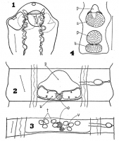

 o, ovary; p, parauterine organ; t, testes; u, uterus; v, vitellarium All figures concern Anonchotaenia rostellata n. sp. They were made with the aid of a camera lucida with details added freehandedly.
Fig. 1. Scolex, dorsal view. The dorsal excretory canals are represented by solid lines, the ventral vessels by double outline. Scale, 150µ. Fig. 2. Partially gravid proglottis, ventral view. Scale, 100µ. Fig. 3. Mature proglottis, ventral view. Scale, 50µ. Fig. 4. Two hindmost proglottides, showing eggs in the parauterine organ of only the terminal segment. Scale, 500µ.
o, ovary; p, parauterine organ; t, testes; u, uterus; v, vitellarium All figures concern Anonchotaenia rostellata n. sp. They were made with the aid of a camera lucida with details added freehandedly.
Fig. 1. Scolex, dorsal view. The dorsal excretory canals are represented by solid lines, the ventral vessels by double outline. Scale, 150µ. Fig. 2. Partially gravid proglottis, ventral view. Scale, 100µ. Fig. 3. Mature proglottis, ventral view. Scale, 50µ. Fig. 4. Two hindmost proglottides, showing eggs in the parauterine organ of only the terminal segment. Scale, 500µ. Best viewed in Firefox


