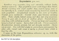
Cestode Scientific Name
| Species ID | 8802 |
|---|---|
| Order | Cyclophyllidea |
| Family | Dilepididae |
| Subfamily | |
| Genus | Eugonodaeum |
| Species | oedicnemi |
| Authority | Beddard, 1913 |
| Taxonomic Status | Valid |
| Valid Name | |
| Synonyms | |
| Genus Record | No |
| Type Species | Yes |
| Verified | No |
| Verified By | |
| Citation(s) |
Beddard, F. E. 1913. Contributions to the anatomy and systematic arrangement of the Cestoidea. XI. On a new tapeworm from dicnemus. Proceedings of the Zoological Society of London 1913(2): 861-877. (5375) Download PDF |
| Redescription | |
| Scientific Name Notes | Type species for genus Eugonodaeum Beddard, 1913 |
Record Data
| Date (MM/DD/YYYY) | Action | User Name |
|---|---|---|
| 02/01/2012 | Created | Salamatin |
| 10/10/2013 | Modified | |
| 05/11/2016 | Modified | B. Barbeau |
| 10/20/2016 | Modified | A. Phillips |
Type Host
| Host Class | Aves | ||||||
|---|---|---|---|---|---|---|---|
| Host Order | Charadriiformes | ||||||
| Host Family | Burhinidae | ||||||
|
Type Host (Literal) |
|
||||||
|
Type Host (Valid) |
|
||||||
| Additional Host(s) | |||||||
| Site in Host | intestine | ||||||
| Host Notes |
Type Locality
| Country | United Kingdom |
|---|---|
| Body of Water | |
| Island(s) | |
| City/Region | London, Zoo (from America) |
| Coordinates | |
| DD Latitude | |
| DD Longitude | |
| Additional Localities | |
| Locality Notes |
Specimens
| Type Material | |
|---|---|
| Total Number of Type Specimens | |
| Voucher Material | |
| Specimen Notes |
Data are given as in original description unless otherwise indicated.
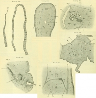
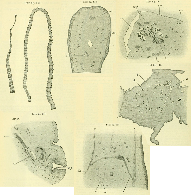 Text-fig. 141. Eugonodaeum oedicnemi. Portions of two examples about twice nat. size. The left-hand figure shows the rather large scolex and whiplash-like anterior part of strobila. The right-hand figure consists of mature segments. Text-fig. 142. Part of a transverse section through a proglottid of Eugonodaeum oedicnemi, to show the arrangement of the water-vascular tubes and of the longitudinal muscles. d. Dorsal vessel. m. Longitudinal muscles. n. Nerve-cord. v. Ventral vessel. Text-fig. 143. Part of a horizontal section through a proglottid of Eugonodaeum oedicnemi. e.s. Egg-sacs. l.v. Ventral water-vascular tube. sp.d. A part of a coil of sperm-duct lying in front of an ovary. t. Testes. t.v. Transverse water-vascular tube. v. Vitelline gland; in front of this is the ovary, of which the darkly stained mature ova are partly received with cavities of the parenchyma. Text-fig. 144. Part of a horizontal section through a proglottid of Eugonodaeum oedicnemi, showing a generative aperture. c. Cirrus extruded from cirrus-sac (shown lying in front of it) and received withing vagina (v.). c.g. Very deep cloaca genitalis. sp.d. A coil of sperm-duct.Text-fig. 145. Part of a horizontal section through a proglottid of Eugonodaeum oedicnemi, to show further course of vagina. e. Egg-sacs. m. Longitudinal muslce-fibres. n. Nerve-cord. V. Vagina, full of sperm and widening above to form a receptaculum seminis, thence bending back again to divide into the two usual branches o. & Vit., which are arranged in the same straight line with each other. Text-fig. 146. A horizontal section through mature proglottid of Eugonodaeum oedicnemi. A. Advanced embryo in egg-holding cavity. B. Less advanced embryo in smaller cavity. v. Ventral water-vascular vessel giving off tranverse vessel.
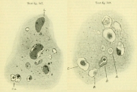
 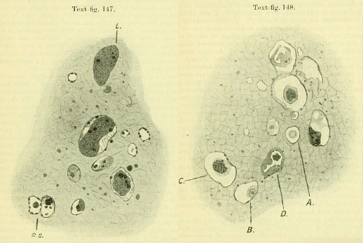 Text-fig. 147. Part of a section illustrated in text-fig. 146 more highly magnified and showing young egg-sacs in parenchyma (e.s.) and testes (t.), which are very much larger. Text-fig. 148. Another part of the same section of Eugonodaeum oedicnemi showing older embryos surrounded by a greater egg-holding cavity in the medullary parenchyma. A, B. Egg surrounded by thick shell and lying in cavity from which nutritive cells (?), such as are shown in es. in text-figure 147, have disappeared. C. Older embryo with large space surrounding it. D. A degenerating (?) egg-holding cavity and embryo.
 Best viewed in Firefox


