Cestode Scientific Name
| Species ID | 8556 |
|---|---|
| Order | Onchoproteocephalidea I |
| Family | Proteocephalidae |
| Subfamily | Gangesiinae |
| Genus | Ritacestus |
| Species | ritaii |
| Authority | (Verma 1926) de Chambrier, Scholz, Ash & Kar, 2011 |
| Taxonomic Status | Valid |
| Valid Name | |
| Synonyms | Proteocephalus ritaii Verma, 1926 |
| Genus Record | No |
| Type Species | Yes |
| Verified | Yes |
| Verified By | T. Scholz |
| Citation(s) |
Verma, S. C. 1926. On a new proteocephalid cestode from an Indian freshwater fish. Allahabad University Studies 2: 353-361. (4137) Download PDFde Chambrier, A., T. Scholz, A. Ash, and P. K. Kar. 2011. Ritacestus gen. n. (Cestoda: Proteocephalidea) and redescription of R. ritaii comb. n., a parasite of Rita rita (Siluriformes) in India. Folia Parasitologica 58(4): 279-288. (5451) Download PDF |
| Redescription |
de Chambrier, A., T. Scholz, A. Ash, and P. K. Kar. 2011. Ritacestus gen. n. (Cestoda: Proteocephalidea) and redescription of R. ritaii comb. n., a parasite of Rita rita (Siluriformes) in India. Folia Parasitologica 58(4): 279-288. (5451) Download PDF |
| Scientific Name Notes |
Record Data
| Date (MM/DD/YYYY) | Action | User Name |
|---|---|---|
| 01/25/2012 | Created | A. Ash, B. Barbeau |
| 06/09/2014 | Modified | |
| 03/18/2016 | Modified | B. Barbeau |
| 04/26/2020 | Modified | T. Scholz |
| 04/28/2020 | Modified | T. Scholz |
| 12/01/2021 | Modified | B. Barbeau |
| 12/03/2021 | Modified | B. Barbeau |
| 06/16/2024 | Modified | T. Scholz |
Type Host
| Host Class | Actinopterygii | ||||||
|---|---|---|---|---|---|---|---|
| Host Order | Siluriformes | ||||||
| Host Family | Bagridae | ||||||
|
Type Host (Literal) |
|
||||||
|
Type Host (Valid) |
|
||||||
| Additional Host(s) | |||||||
| Site in Host | anterior intestine | ||||||
| Host Notes |
Type Locality
| Country | India |
|---|---|
| Body of Water | Ganges and Jumna rivers |
| Island(s) | |
| City/Region | Northern India |
| Coordinates | |
| DD Latitude | |
| DD Longitude | |
| Additional Localities | |
| Locality Notes |
Specimens
| Type Material | - MHNG-PLAT No. 63242 (neotype) |
|---|---|
| Total Number of Type Specimens | Based on 7 specimens from West Bengal, India |
| Voucher Material | |
| Specimen Notes |
Data are given as in original description unless otherwise indicated.
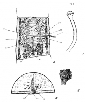

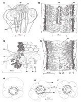
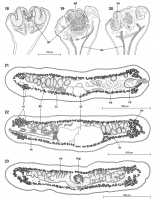
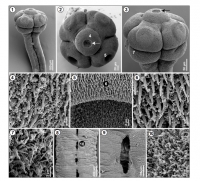
 Plate 1. Fig. 1Anterior end of P. ritaii in outline, showing Scolex with Suckers and Neck. X 30. Fig. 2Scolex mounted entire, showing the Four Suckers with marginal flaps round the Apertures; the Apical Organ, and the Grooves between the Suckers. X 45. Fig. 3A Mature Proglottid. X 25. Fig. 4Part of Horizontal Section showing Genital Atrium, and the Cirrus Sac and Vagina Openings. X 40.
 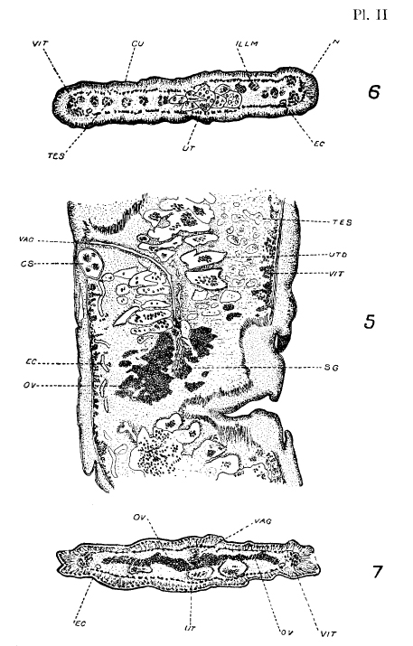 Plate 2. Fig. 5Horizontal Section (slightly oblique) of a Mature Proglottid. X 40. Fig. 6Transverse Section of a Mature Proglottid anterior to the Genital Openings. X 50. Fig. 7Transverse Section of a Mature Proglottid in the region of the Ovarian Isthmus. X 50.
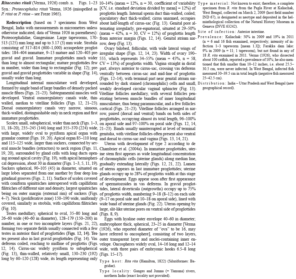
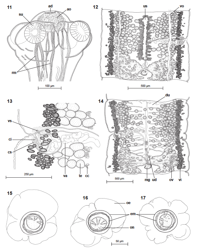 Figs. 1117. Ritacestus ritaii (Verma, 1926) comb. n. from Rita rita, India. Fig. 11. Scolex, dorsoventral view (neotype - INVE 63242). Fig. 12. Mature proglottis, ventral view (IPCAS C603). Fig. 13. Terminal genitalia, dorsal view (IPCAS C603). Fig. 14. Gravid proglottis, dorsal view (IPCAS C603). Figs. 1517. Eggs drawn in distilled water. Abbreviations: ad - apical depression; ao - apical organ; cc - chromophilic cells on the apex of diverticula of uterus; ci - cirrus; cs - cirrus-sac; em - embryophore; mg - Mehlis' gland; oe - outer envelope; on - oncosphere; ov - ovary; rm - retractor muscles; su - suckers; te - testes; ud - uteroduct; us - uterine stem; va - vas deferens; vi - vitelline follicles; vo - ventral osmoregulatory canal; vs - vaginal sphincter.
  Figs. 1823. Ritacestus ritaii (Verma, 1926) comb. n. from Rita rita, India. Figs. 1820. Longitudinal sections of the scolex (INVE 78789). Figs. 21-23. Cross sections at level of cirrus-sac, uterus and overy, respectively (INVE 78786). Abbreviations: ad - apical depression; cc - chromophilic cells on the apex of diverticula of uterus; cs - cirrus-sac; gc - glandular cells; lm - internal longitudinal musculature; mg - Mehlis' glad; ov - overy; rm - retractor muscles; su - suckers; te - testes; us - uterus; va - vas deferens; vc - vaginal canal; vi - vitelline follicles; vo - ventral osmoregulatory canal (anastomosed).
  Figs. 1-10. Ritacestus ritaii (Verma, 1926) comb. n. from Rita rita, India. Scanning electron micrographs. Fig. 1. Scolex and neck region, dorsoventral view. Fig. 2. Scolex, apical view; note an apical hemispherical depression (arrow). Fig. 3. Scolex, dorsventral vieew; note an apical hemispherical depression (arrow). Figs. 4-7. Microtriches (coniform spinitriches interspersed with capilliform filitriches) on the surface of apical organ (4), sucker cavity (5), external rim of suckers (6) and lobes of anterior part of neck (7), respectively. Figs. 8,9. Uterine pores. Fig. 10. Microtriches (capilliform filitriches) on strobila. Note: small numbers in Figs. 2, 3, 5 and 8 correspond to the figures showing higher magnification images of marked regions.
Best viewed in Firefox


