Line Drawing 1
Figs 16. Acanthobothrium bobconniorum, sp. nov. 1, Scolex. 2, Hooks. 3, Sub-terminal proglottid. Arrow indicates location
of cross section shown in Fig. 6. 4, Terminal proglottid. 5, Whole worm. 6, ... MoreFigs 16. Acanthobothrium bobconniorum, sp. nov. 1, Scolex. 2, Hooks. 3, Sub-terminal proglottid. Arrow indicates location
of cross section shown in Fig. 6. 4, Terminal proglottid. 5, Whole worm. 6, Cross section through sub-terminal proglottid at level
of ovary. Abbreviations: O, ovary; T, testis; U, uterus; V, vitelline follicle; VA, vagina; VED, ventral excretory duct. |
Line Drawing 2
|
Photo Micrograph
|
Scanning Electron Micrograph
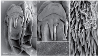
Figs 1827. Scanning electron micrographs of Acanthobothrium bobconniorum, 1822, Acanthobothrium bobconniorum, sp. nov. 18, Scolex. 19, Detail of muscular pad and hooks. 20, Distal bothridial surface... MoreFigs 1827. Scanning electron micrographs of Acanthobothrium bobconniorum, 1822, Acanthobothrium bobconniorum, sp. nov. 18, Scolex. 19, Detail of muscular pad and hooks. 20, Distal bothridial surface. 21, Proximal bothridial surface at rim of bothridium. 22, Surface of cephalic peduncle. Small white numbers indicate locations of Figs 2022. |
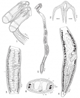
 Figs 16. Acanthobothrium bobconniorum, sp. nov. 1, Scolex. 2, Hooks. 3, Sub-terminal proglottid. Arrow indicates location
of cross section shown in Fig. 6. 4, Terminal proglottid. 5, Whole worm. 6, Cross section through sub-terminal proglottid at level
of ovary. Abbreviations: O, ovary; T, testis; U, uterus; V, vitelline follicle; VA, vagina; VED, ventral excretory duct.
Figs 16. Acanthobothrium bobconniorum, sp. nov. 1, Scolex. 2, Hooks. 3, Sub-terminal proglottid. Arrow indicates location
of cross section shown in Fig. 6. 4, Terminal proglottid. 5, Whole worm. 6, Cross section through sub-terminal proglottid at level
of ovary. Abbreviations: O, ovary; T, testis; U, uterus; V, vitelline follicle; VA, vagina; VED, ventral excretory duct. 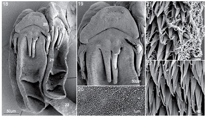 Figs 1827. Scanning electron micrographs of Acanthobothrium bobconniorum, 1822, Acanthobothrium bobconniorum, sp. nov. 18, Scolex. 19, Detail of muscular pad and hooks. 20, Distal bothridial surface. 21, Proximal bothridial surface at rim of bothridium. 22, Surface of cephalic peduncle. Small white numbers indicate locations of Figs 2022.