Line Drawing 1
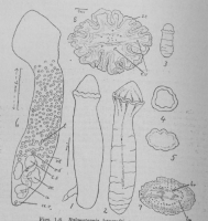
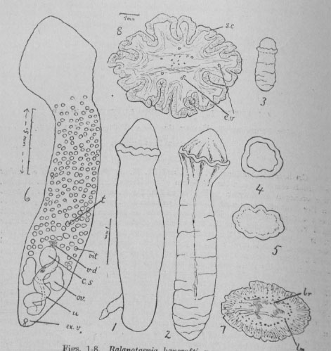
Figs. 1-8. balanotaenia bancrofti, n.gen. et. sp. 1. Lateral view of adult with extruded cirrus. 2. Ventral view of adult. 3. Smallest specimen observed, 1.1 mm. by .4 mm. (in formalin). 4, 5. End vie... MoreFigs. 1-8. balanotaenia bancrofti, n.gen. et. sp. 1. Lateral view of adult with extruded cirrus. 2. Ventral view of adult. 3. Smallest specimen observed, 1.1 mm. by .4 mm. (in formalin). 4, 5. End views of scolices (in formalin). 6. General view (dorsal) ; stained preparation, somewhat compressed. 7. Transverse section of anterior part of scolex in region of brain. 8. Transverse section in region of "frill" ; note deeply folded surface.
Figs. 1 to 5 have been drawn to the scale indicated beside Fig. 1. Figs. 7 and 8 drawn to scale beside Fig. 8. All figures have been drawn with the aid of a camera lucida.
References to lettering:--br., brain; c.s., cirrus sac; cu., cuticle; e., egg; g.u., glandular uterus; l.m., longitudinal muscles; m, muscles; n., nerve; od., oviduct; o.i., ovarian isthmus; ov., ovary; r.s., receptaculum seminis; s.c., subcuticula; s.g., shell gland; t., testis; t.m., transverse muscle fibers; u., uterus; u.a., uterine opening into genital atrium; u.c., uterine cavity; u.d., uterine duct; v., vagina; v.d., vas deferens or vesicula seminalis; vit., vitelline follicle; vit. d., vitelline duct. |
Line Drawing 2
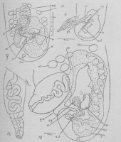
Figs. 9-12. Balanotaenia bancrofti, n.ge. et sp. 9. Posterior end, showing anatomy, viewed ventrally and slightly obliquely: shell gland, vitelline duct, oviduct and also the testes and vitellaria in ... MoreFigs. 9-12. Balanotaenia bancrofti, n.ge. et sp. 9. Posterior end, showing anatomy, viewed ventrally and slightly obliquely: shell gland, vitelline duct, oviduct and also the testes and vitellaria in the vicinity of uterus and cirrus sac are omitted. 10. Lateral views of posterior region, examined in clove oil; showing partly extruded cirrus; outline of ovary and position of commencement of oviduct indicated by broken lines: position of shell gland marked by dots surrounding a shaded area: the thick walls and sinous lumen of the uterus are indicated. Most of the vitellaria have been omitted. 11. Posterior end of a specimen examined in clove oil: cirrus and sac almost fully extended. Lateral view. 12. Extruded cirrus and sac of specimen figured in Fig. 11-drawn to same scale as Fig. 10. |
Photo Micrograph
|
Scanning Electron Micrograph
|

 Figs. 1-8. balanotaenia bancrofti, n.gen. et. sp. 1. Lateral view of adult with extruded cirrus. 2. Ventral view of adult. 3. Smallest specimen observed, 1.1 mm. by .4 mm. (in formalin). 4, 5. End views of scolices (in formalin). 6. General view (dorsal) ; stained preparation, somewhat compressed. 7. Transverse section of anterior part of scolex in region of brain. 8. Transverse section in region of "frill" ; note deeply folded surface.
Figs. 1 to 5 have been drawn to the scale indicated beside Fig. 1. Figs. 7 and 8 drawn to scale beside Fig. 8. All figures have been drawn with the aid of a camera lucida.
References to lettering:--br., brain; c.s., cirrus sac; cu., cuticle; e., egg; g.u., glandular uterus; l.m., longitudinal muscles; m, muscles; n., nerve; od., oviduct; o.i., ovarian isthmus; ov., ovary; r.s., receptaculum seminis; s.c., subcuticula; s.g., shell gland; t., testis; t.m., transverse muscle fibers; u., uterus; u.a., uterine opening into genital atrium; u.c., uterine cavity; u.d., uterine duct; v., vagina; v.d., vas deferens or vesicula seminalis; vit., vitelline follicle; vit. d., vitelline duct.
Figs. 1-8. balanotaenia bancrofti, n.gen. et. sp. 1. Lateral view of adult with extruded cirrus. 2. Ventral view of adult. 3. Smallest specimen observed, 1.1 mm. by .4 mm. (in formalin). 4, 5. End views of scolices (in formalin). 6. General view (dorsal) ; stained preparation, somewhat compressed. 7. Transverse section of anterior part of scolex in region of brain. 8. Transverse section in region of "frill" ; note deeply folded surface.
Figs. 1 to 5 have been drawn to the scale indicated beside Fig. 1. Figs. 7 and 8 drawn to scale beside Fig. 8. All figures have been drawn with the aid of a camera lucida.
References to lettering:--br., brain; c.s., cirrus sac; cu., cuticle; e., egg; g.u., glandular uterus; l.m., longitudinal muscles; m, muscles; n., nerve; od., oviduct; o.i., ovarian isthmus; ov., ovary; r.s., receptaculum seminis; s.c., subcuticula; s.g., shell gland; t., testis; t.m., transverse muscle fibers; u., uterus; u.a., uterine opening into genital atrium; u.c., uterine cavity; u.d., uterine duct; v., vagina; v.d., vas deferens or vesicula seminalis; vit., vitelline follicle; vit. d., vitelline duct. 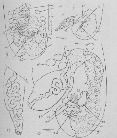 Figs. 9-12. Balanotaenia bancrofti, n.ge. et sp. 9. Posterior end, showing anatomy, viewed ventrally and slightly obliquely: shell gland, vitelline duct, oviduct and also the testes and vitellaria in the vicinity of uterus and cirrus sac are omitted. 10. Lateral views of posterior region, examined in clove oil; showing partly extruded cirrus; outline of ovary and position of commencement of oviduct indicated by broken lines: position of shell gland marked by dots surrounding a shaded area: the thick walls and sinous lumen of the uterus are indicated. Most of the vitellaria have been omitted. 11. Posterior end of a specimen examined in clove oil: cirrus and sac almost fully extended. Lateral view. 12. Extruded cirrus and sac of specimen figured in Fig. 11-drawn to same scale as Fig. 10.

