Line Drawing 1
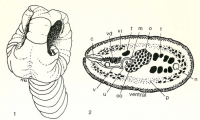
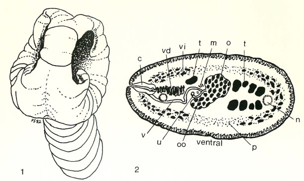
Abbreviations : c, cirrus; m, Mehlis gland; n, nerve cord; o, ovary, oo, ootype; p, parenchymal longitudinal muscles; t, testes; u, uterus; v, vagina; vd, vas deferens; vi, vitellaria.
Fig. 1. Diagra... MoreAbbreviations : c, cirrus; m, Mehlis gland; n, nerve cord; o, ovary, oo, ootype; p, parenchymal longitudinal muscles; t, testes; u, uterus; v, vagina; vd, vas deferens; vi, vitellaria.
Fig. 1. Diagram of scolex showing median groove, apical disk, and muscular bothria. X64.
Fig. 2. Diagram of transverse section of the segment at level of the ovary and cirrus sac. X64. |
Line Drawing 2
|
Photo Micrograph
|
Scanning Electron Micrograph
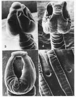
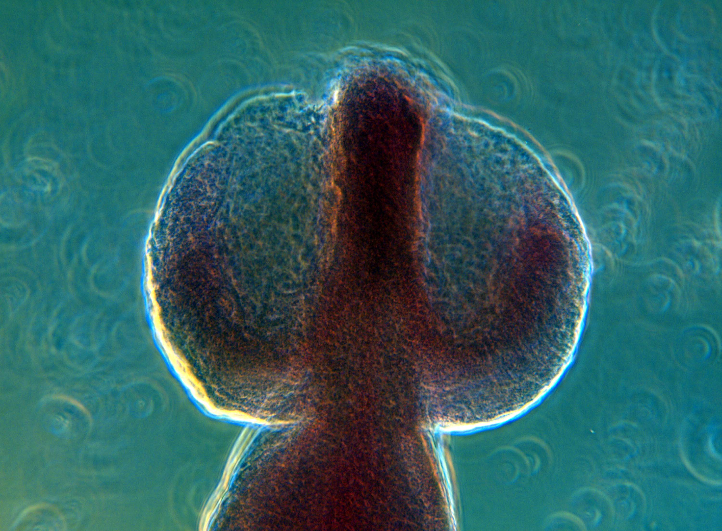
Fig. 3. SEM photomicrograph, lateral view of scolex showing apical disk and median groove. X86.
Fig. 4. SEM photomicrograph, lateral view of scolex. X72. Fig. 5. SEM photomicrograph, dorsal view of ... MoreFig. 3. SEM photomicrograph, lateral view of scolex showing apical disk and median groove. X86.
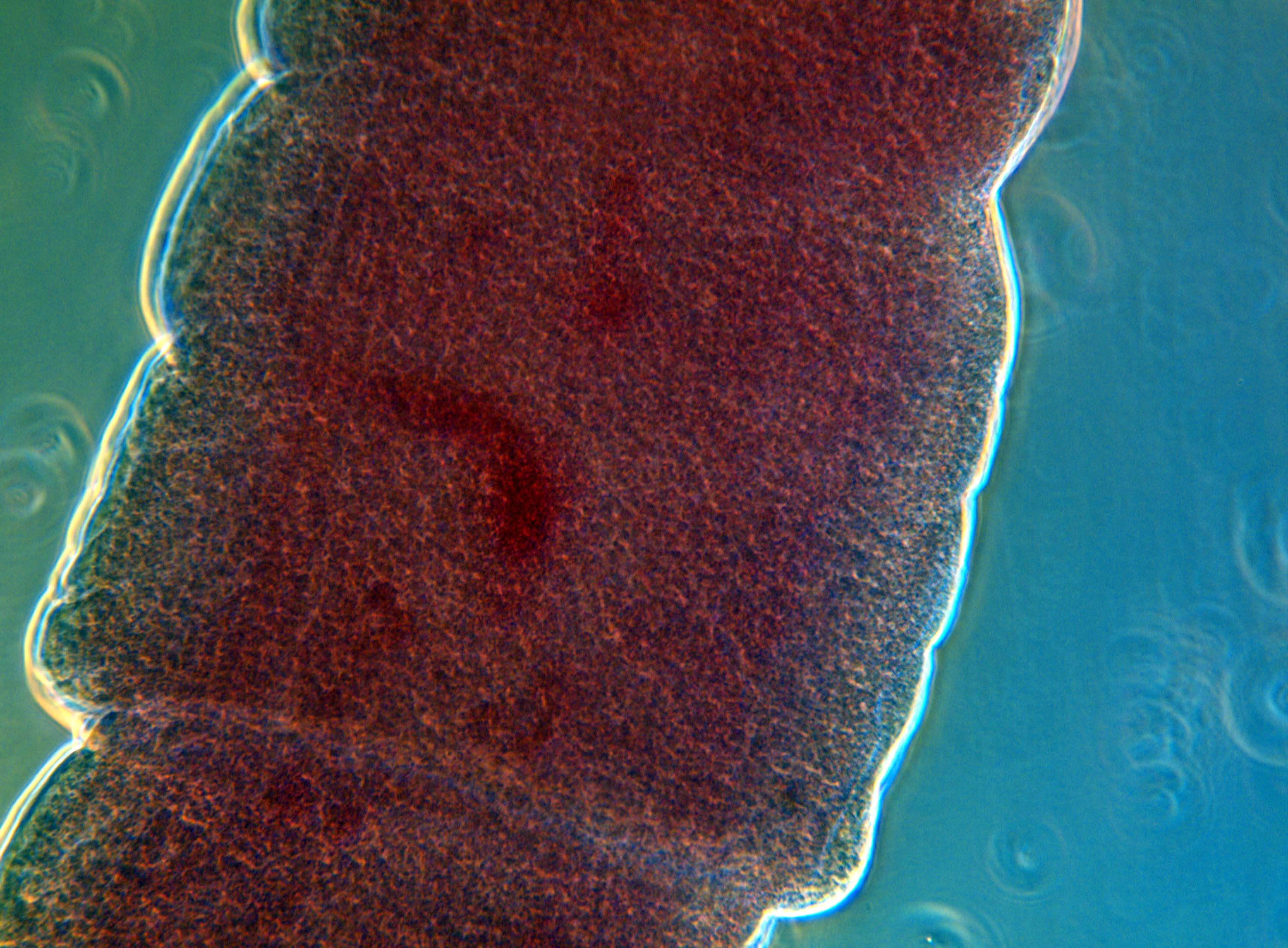
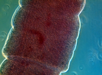
Fig. 4. SEM photomicrograph, lateral view of scolex. X72. Fig. 5. SEM photomicrograph, dorsal view of bothrium and apical disk. X90. Fig. 6. Segment with secondary segmentation and prominent genital pore. X100. |

 Abbreviations : c, cirrus; m, Mehlis gland; n, nerve cord; o, ovary, oo, ootype; p, parenchymal longitudinal muscles; t, testes; u, uterus; v, vagina; vd, vas deferens; vi, vitellaria.
Fig. 1. Diagram of scolex showing median groove, apical disk, and muscular bothria. X64.
Fig. 2. Diagram of transverse section of the segment at level of the ovary and cirrus sac. X64.
Abbreviations : c, cirrus; m, Mehlis gland; n, nerve cord; o, ovary, oo, ootype; p, parenchymal longitudinal muscles; t, testes; u, uterus; v, vagina; vd, vas deferens; vi, vitellaria.
Fig. 1. Diagram of scolex showing median groove, apical disk, and muscular bothria. X64.
Fig. 2. Diagram of transverse section of the segment at level of the ovary and cirrus sac. X64.  Fig. 3. SEM photomicrograph, lateral view of scolex showing apical disk and median groove. X86.
Fig. 4. SEM photomicrograph, lateral view of scolex. X72. Fig. 5. SEM photomicrograph, dorsal view of bothrium and apical disk. X90. Fig. 6. Segment with secondary segmentation and prominent genital pore. X100.
Fig. 3. SEM photomicrograph, lateral view of scolex showing apical disk and median groove. X86.
Fig. 4. SEM photomicrograph, lateral view of scolex. X72. Fig. 5. SEM photomicrograph, dorsal view of bothrium and apical disk. X90. Fig. 6. Segment with secondary segmentation and prominent genital pore. X100.