Line Drawing 1
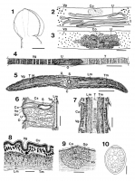
Figs. 1-10. Diphyllobothrium orcini n. sp. from killer whale, Orcinus orca (Linnaeus, 1758). Fig. 1. Scolex, lateral view (Scale bar = 0.2mm). Fig. 2. Uterine loops of mature segment (about 35cm behin... MoreFigs. 1-10. Diphyllobothrium orcini n. sp. from killer whale, Orcinus orca (Linnaeus, 1758). Fig. 1. Scolex, lateral view (Scale bar = 0.2mm). Fig. 2. Uterine loops of mature segment (about 35cm behind the scolex). ventral view (Scale bar = 0.2mm). Fig. 3. Uterine loops of gravid segment, ventral view (Scale bar = 1.0mm). Fig. 4. Gravid segment (about 90cm behind the scolex), ventral view (Scale bar = 2.0mm). Fig. 5. Transverse section of gravid segment passing through ihe level of the genital opening (Scale bar = 2.0mm). Fig. 6. Sagittal section of gravid segment passing through the genital field (Scale bar = 0.3mm). Fig. 7. Sagittal section of gravid segment passing through the lateral field (Scale bar = 0.5mm). Fig. 8. Transverse section of gravid segment, showing details of ihe dorsal musculature (Scale bar - 0.2mm). Fig. 9. Cirrus opening and genital papillae of gravid segment, ventral view (Scale bar = 0.4mm). Fig. 10. Egg (Scale bar = 0.02mm).
C: cirrus, Cs: cirrus-sac, Co: Cirrus opening, Cu: cuticle, Gp: genital papillae, Lm: longitudinal muscle layer, N: nerve trunk, O: ovary, S: seminal vesicle, T: testis, Tm: transverse muscle layer, U: uterus, Uo: uterine opening, Vg: vitellinc gland, Vo: vaginal opening. |
Line Drawing 2
|
Photo Micrograph
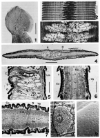
Photos. 1-9. Diphyllobolftrium orcini n. sp. from killer whale, Orcinus orca (Linnaeus, 1758). Photo. I. Scolex, lateral view (Scale bar = 0.2mm). Photo. 2. Gravid segment (about 60cm behind the scole... MorePhotos. 1-9. Diphyllobolftrium orcini n. sp. from killer whale, Orcinus orca (Linnaeus, 1758). Photo. I. Scolex, lateral view (Scale bar = 0.2mm). Photo. 2. Gravid segment (about 60cm behind the scolex), ventral view (Scale bar = 2.0mm). Photo. 3. High-magnification ul Plioto. 1, showing details of uterine loops (Scale bar 0.5mm). Photo. 4. Transverse section of gravid segment (Scale bar = 2.0mm). Photo. 5. Sagittal section of gravid segment passing through the genital field (Scale bar = 0.3mm). Photo. 6. Sagittal section of gravid segment passing through the lateral field (Scale bar = 0.5mm). Photo. 7. Transverse section of gravid segment, showing details of the lateral field (Scale bar = 0.4mm). Photo. 8. Genital papillae and cirrus opening of gravid segment by scanning electron microscopy (Scale bar = 0.2mm). Photo. 9. Egg-shell surface by scanning electron microscopy (Scale bar = 2.0 um).
C: cirrus, Cs: cirrus-sac, Co: Cirrus opening, Cu: cuticle, Gp: genital papillae, Lm: longitudinal muscle layer, N: nerve trunk, O: ovary, S: seminal vesicle, T: testis, Tm: transverse muscle layer, U: uterus, Uo: uterine opening, Vg: vitellinc gland, Vo: vaginal opening. |
Scanning Electron Micrograph
|
 Figs. 1-10. Diphyllobothrium orcini n. sp. from killer whale, Orcinus orca (Linnaeus, 1758). Fig. 1. Scolex, lateral view (Scale bar = 0.2mm). Fig. 2. Uterine loops of mature segment (about 35cm behind the scolex). ventral view (Scale bar = 0.2mm). Fig. 3. Uterine loops of gravid segment, ventral view (Scale bar = 1.0mm). Fig. 4. Gravid segment (about 90cm behind the scolex), ventral view (Scale bar = 2.0mm). Fig. 5. Transverse section of gravid segment passing through ihe level of the genital opening (Scale bar = 2.0mm). Fig. 6. Sagittal section of gravid segment passing through the genital field (Scale bar = 0.3mm). Fig. 7. Sagittal section of gravid segment passing through the lateral field (Scale bar = 0.5mm). Fig. 8. Transverse section of gravid segment, showing details of ihe dorsal musculature (Scale bar - 0.2mm). Fig. 9. Cirrus opening and genital papillae of gravid segment, ventral view (Scale bar = 0.4mm). Fig. 10. Egg (Scale bar = 0.02mm).
C: cirrus, Cs: cirrus-sac, Co: Cirrus opening, Cu: cuticle, Gp: genital papillae, Lm: longitudinal muscle layer, N: nerve trunk, O: ovary, S: seminal vesicle, T: testis, Tm: transverse muscle layer, U: uterus, Uo: uterine opening, Vg: vitellinc gland, Vo: vaginal opening.
Figs. 1-10. Diphyllobothrium orcini n. sp. from killer whale, Orcinus orca (Linnaeus, 1758). Fig. 1. Scolex, lateral view (Scale bar = 0.2mm). Fig. 2. Uterine loops of mature segment (about 35cm behind the scolex). ventral view (Scale bar = 0.2mm). Fig. 3. Uterine loops of gravid segment, ventral view (Scale bar = 1.0mm). Fig. 4. Gravid segment (about 90cm behind the scolex), ventral view (Scale bar = 2.0mm). Fig. 5. Transverse section of gravid segment passing through ihe level of the genital opening (Scale bar = 2.0mm). Fig. 6. Sagittal section of gravid segment passing through the genital field (Scale bar = 0.3mm). Fig. 7. Sagittal section of gravid segment passing through the lateral field (Scale bar = 0.5mm). Fig. 8. Transverse section of gravid segment, showing details of ihe dorsal musculature (Scale bar - 0.2mm). Fig. 9. Cirrus opening and genital papillae of gravid segment, ventral view (Scale bar = 0.4mm). Fig. 10. Egg (Scale bar = 0.02mm).
C: cirrus, Cs: cirrus-sac, Co: Cirrus opening, Cu: cuticle, Gp: genital papillae, Lm: longitudinal muscle layer, N: nerve trunk, O: ovary, S: seminal vesicle, T: testis, Tm: transverse muscle layer, U: uterus, Uo: uterine opening, Vg: vitellinc gland, Vo: vaginal opening.  Photos. 1-9. Diphyllobolftrium orcini n. sp. from killer whale, Orcinus orca (Linnaeus, 1758). Photo. I. Scolex, lateral view (Scale bar = 0.2mm). Photo. 2. Gravid segment (about 60cm behind the scolex), ventral view (Scale bar = 2.0mm). Photo. 3. High-magnification ul Plioto. 1, showing details of uterine loops (Scale bar 0.5mm). Photo. 4. Transverse section of gravid segment (Scale bar = 2.0mm). Photo. 5. Sagittal section of gravid segment passing through the genital field (Scale bar = 0.3mm). Photo. 6. Sagittal section of gravid segment passing through the lateral field (Scale bar = 0.5mm). Photo. 7. Transverse section of gravid segment, showing details of the lateral field (Scale bar = 0.4mm). Photo. 8. Genital papillae and cirrus opening of gravid segment by scanning electron microscopy (Scale bar = 0.2mm). Photo. 9. Egg-shell surface by scanning electron microscopy (Scale bar = 2.0 um).
C: cirrus, Cs: cirrus-sac, Co: Cirrus opening, Cu: cuticle, Gp: genital papillae, Lm: longitudinal muscle layer, N: nerve trunk, O: ovary, S: seminal vesicle, T: testis, Tm: transverse muscle layer, U: uterus, Uo: uterine opening, Vg: vitellinc gland, Vo: vaginal opening.
Photos. 1-9. Diphyllobolftrium orcini n. sp. from killer whale, Orcinus orca (Linnaeus, 1758). Photo. I. Scolex, lateral view (Scale bar = 0.2mm). Photo. 2. Gravid segment (about 60cm behind the scolex), ventral view (Scale bar = 2.0mm). Photo. 3. High-magnification ul Plioto. 1, showing details of uterine loops (Scale bar 0.5mm). Photo. 4. Transverse section of gravid segment (Scale bar = 2.0mm). Photo. 5. Sagittal section of gravid segment passing through the genital field (Scale bar = 0.3mm). Photo. 6. Sagittal section of gravid segment passing through the lateral field (Scale bar = 0.5mm). Photo. 7. Transverse section of gravid segment, showing details of the lateral field (Scale bar = 0.4mm). Photo. 8. Genital papillae and cirrus opening of gravid segment by scanning electron microscopy (Scale bar = 0.2mm). Photo. 9. Egg-shell surface by scanning electron microscopy (Scale bar = 2.0 um).
C: cirrus, Cs: cirrus-sac, Co: Cirrus opening, Cu: cuticle, Gp: genital papillae, Lm: longitudinal muscle layer, N: nerve trunk, O: ovary, S: seminal vesicle, T: testis, Tm: transverse muscle layer, U: uterus, Uo: uterine opening, Vg: vitellinc gland, Vo: vaginal opening. 

