Line Drawing 1
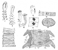
Fig. 7. Head and anterior segments; 12x. Fig. 8. Same specimen, lateral view; 12x. Fig. 9. Another specimen, marginal view, corresponding to lateral margin of body; 12x. Fig. 10. Anterior view of same... MoreFig. 7. Head and anterior segments; 12x. Fig. 8. Same specimen, lateral view; 12x. Fig. 9. Another specimen, marginal view, corresponding to lateral margin of body; 12x. Fig. 10. Anterior view of same specimen; 12x. Fig. 11. Front view of head; 12x. Fig. 12. Antero-median segments; 12x. Fig. 13.Postero-median segments; 12x. Fig. 14. Posterior segments; 12x. Fig. 15. Longitudinal ventral section. Zeiss 2/A, draw-tube open, vg, inner viteline gland. |
Line Drawing 2
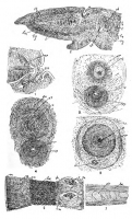
Fig. 1. Genital cloaca with external end og cirrus-pouch and vagina spincter, from longitudinal, ventral section. Zeiss 2/A, draw-tube closed. Fig. 2. Part of transverse section of body through cirrus... MoreFig. 1. Genital cloaca with external end og cirrus-pouch and vagina spincter, from longitudinal, ventral section. Zeiss 2/A, draw-tube closed. Fig. 2. Part of transverse section of body through cirrus-pouch and external, dorsal, opening of uterus. Zeiss 2/A, draw-tube closed. Fig. 3. Section of cirrus and cirrus-pouch and vaginal sphincter, near ventral surface, from longitudinal, horizontal section of body. Zeiss 2/A, draw-tube open. Fig. 4. Same, section made a little deeper in body, lm, longitudinal muscles of cirrus-bulb, Fig. 3. Zeiss 2/A, draw-tube open. Fig. 5. Section of cirrus and cirrus-pouch and vagina, from longitudinal, horizontal section of body, not so much magnified as 3 and 4. Zeiss 2/A, draw-tube closed. Fig. 6. Portion of transverse section. Zeiss 2/A, draw-tube closed. Fig. 7. Longitudinal muscles and calcereous bodies. Zeiss 2/A, draw-tube open. |
Photo Micrograph
|
Scanning Electron Micrograph
|
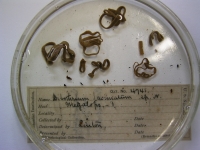

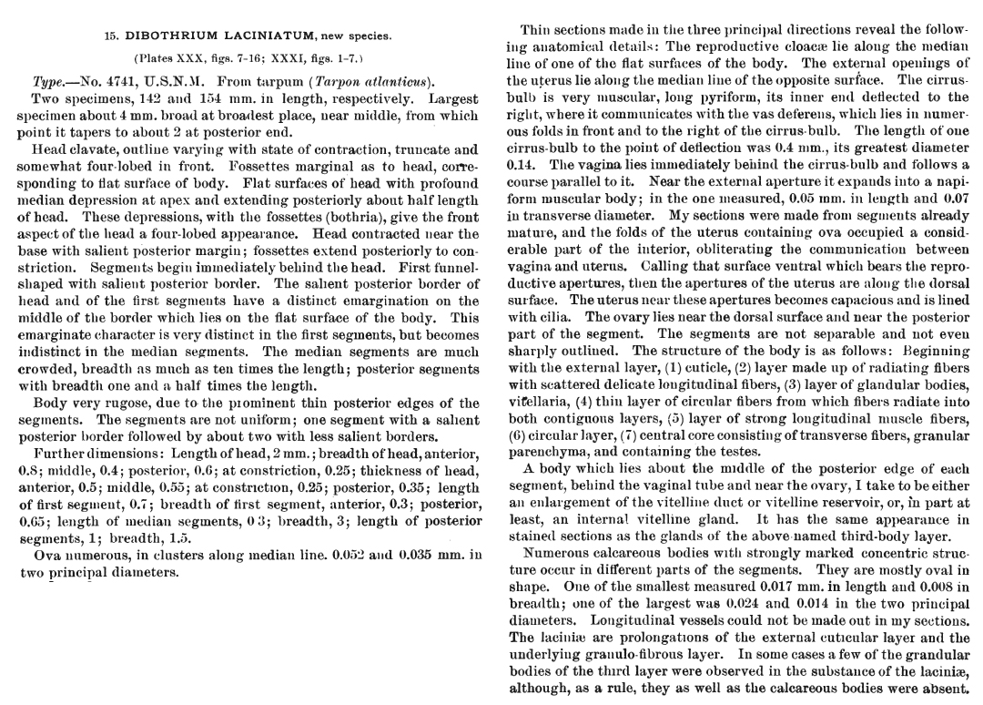
 Fig. 7. Head and anterior segments; 12x. Fig. 8. Same specimen, lateral view; 12x. Fig. 9. Another specimen, marginal view, corresponding to lateral margin of body; 12x. Fig. 10. Anterior view of same specimen; 12x. Fig. 11. Front view of head; 12x. Fig. 12. Antero-median segments; 12x. Fig. 13.Postero-median segments; 12x. Fig. 14. Posterior segments; 12x. Fig. 15. Longitudinal ventral section. Zeiss 2/A, draw-tube open, vg, inner viteline gland.
 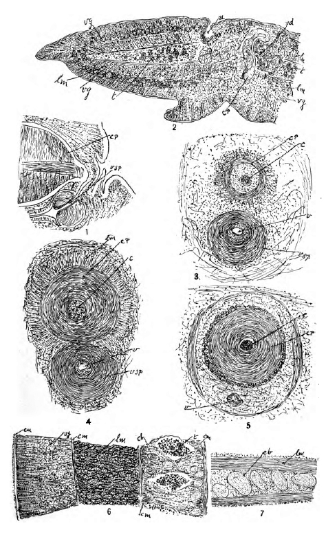 Fig. 1. Genital cloaca with external end og cirrus-pouch and vagina spincter, from longitudinal, ventral section. Zeiss 2/A, draw-tube closed. Fig. 2. Part of transverse section of body through cirrus-pouch and external, dorsal, opening of uterus. Zeiss 2/A, draw-tube closed. Fig. 3. Section of cirrus and cirrus-pouch and vaginal sphincter, near ventral surface, from longitudinal, horizontal section of body. Zeiss 2/A, draw-tube open. Fig. 4. Same, section made a little deeper in body, lm, longitudinal muscles of cirrus-bulb, Fig. 3. Zeiss 2/A, draw-tube open. Fig. 5. Section of cirrus and cirrus-pouch and vagina, from longitudinal, horizontal section of body, not so much magnified as 3 and 4. Zeiss 2/A, draw-tube closed. Fig. 6. Portion of transverse section. Zeiss 2/A, draw-tube closed. Fig. 7. Longitudinal muscles and calcereous bodies. Zeiss 2/A, draw-tube open.
 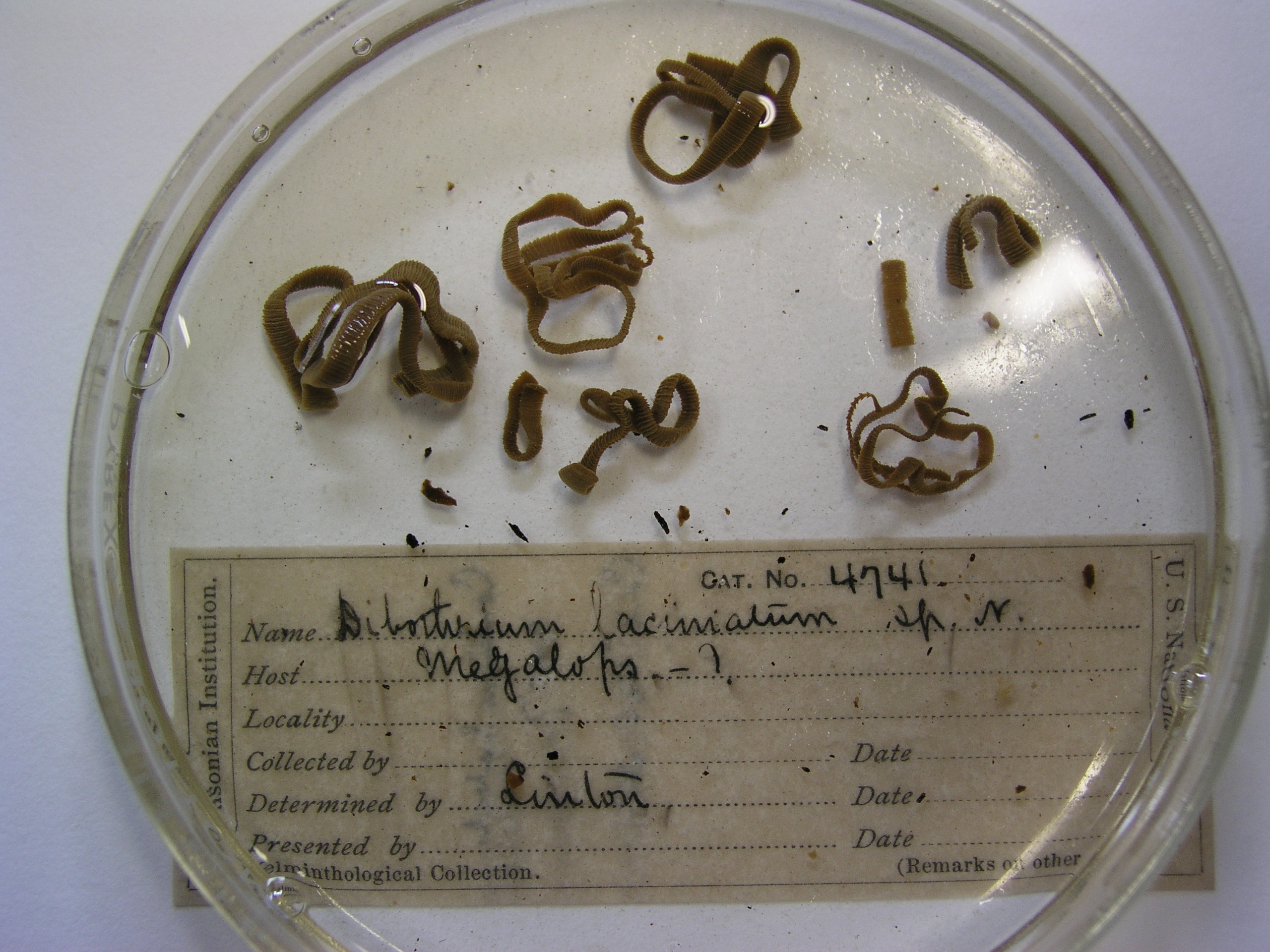 -USNPC No. 4741 (holotype)
 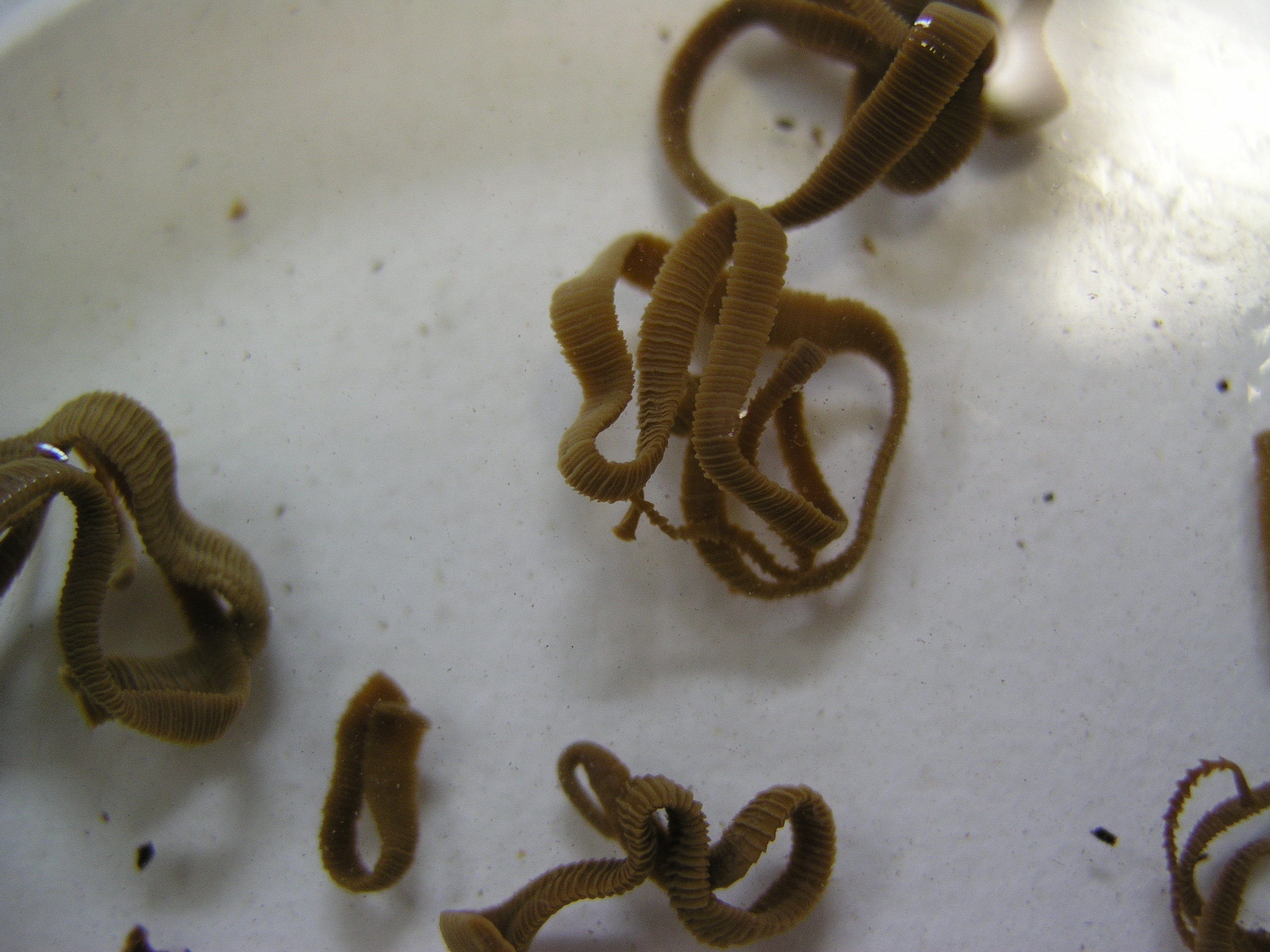 -USNPC No. 4741 (holotype)

