Line Drawing 1
EXPLANATION OF PLATES I AND II
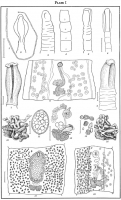
All figures are camera lucida drawings unless otherwise stated. The projected scale for figures 9, 12, 15, I6, i8, I9, 2I, 22, 24, and 28=0.20 mm.; for figures 6, 13, 2... MoreEXPLANATION OF PLATES I AND II
All figures are camera lucida drawings unless otherwise stated. The projected scale for figures 9, 12, 15, I6, i8, I9, 2I, 22, 24, and 28=0.20 mm.; for figures 6, 13, 23, 25, and 29=0.05 mm.; for figure I=30 mm.; for figures 7 and 17=O.I5 mm.; for figure II= 0.3 mm.
Fig. I.-The entire worm.
Figs. 2, 3, and 5.-Free-hand sketches of the scolex in successive movements.
Fig. 4.-Anterior segments, dorsal view, showing position of hermaphroditic pore.
Fig. 6.-Lateral view, toto mount of scolex. Bouin's fixation.
Fig. 7.-Surficial view of scolex and terminal disc, toto mount.
Fig. 8.-Early developing proglottid showing in ventral view testes, excretory ducts, genital complex, uterine tube and pore.
Fig. 9.-Scolex, toto mount, showing end view terminal disc.
Fig. 10.-Wax reconstruction, genital complex seen in anterior ventral oblique view.
Fig. 11.-Segmenting operculate egg some time after laying.
Fig. 12.-Surficial view genital complex, toto mount. Hermaphroditic pore dorsal.
Fig. 13.-Calcareous bodies from cortical layer.
Fig. 14.-Genital complex, wax reconstruction, dorsal view.
Fig. 15.-Mature proglottid, toto mount, ventral view.
Fig. 16.-Proglottid more anterior in segments, ventral view. |
Line Drawing 2
EXPLANATION OF PLATES I AND II
All figures are camera lucida drawings unless otherwise stated. The projected scale for figures 9, 12, 15, I6, i8, I9, 2I, 22, 24, and 28=0.20 mm.; for figures 6, 13, 2... MoreEXPLANATION OF PLATES I AND II
All figures are camera lucida drawings unless otherwise stated. The projected scale for figures 9, 12, 15, I6, i8, I9, 2I, 22, 24, and 28=0.20 mm.; for figures 6, 13, 23, 25, and 29=0.05 mm.; for figure I=30 mm.; for figures 7 and 17=O.I5 mm.; for figure II= 0.3 mm.
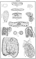
Fig. 17.-Cross-section through bothria showing four main excretory ducts.
Figs. 18 and 19.-Cross-section through proglottids in region of uterine pore and sac.
Fig. 20.-Surface view of cuticula from scolex; oil immersion.
Fig. 21.-Sagittal section through scolex, showing distribution of ganglionic masses; portions of excretory ducts and nerve trunks.
Fig. 22.-Longitudinal section, cirrus sac and vagina.
Fig. 23.-Section through oocapt, oviduct, and ovary.
Fig. 24.-Cross-section of proglottid, showing distribution of muscle fibers, ovary, testes, excretory ducts, vitelline duct, nerve cords, and vitellaria.
Fig. 25.-Longitudinal section near cortical medulla, with a testis, excretory duct, muscles, vitellaria, and two layers of cuticula showing.
Fig. 26.-An ovum (left), spermatozoon (center), and vitelline cells (right).
Fig. 27.-Sagittal section through vitelline reservoir, at juncture with ootype and fertilization tube.
Fig. 28.-Sagittal section through entire proglottid, ventral surface.
Fig. 29.-Section through cloacal region and cirrus sac. |
Photo Micrograph
|
Scanning Electron Micrograph
|


 EXPLANATION OF PLATES I AND II
All figures are camera lucida drawings unless otherwise stated. The projected scale for figures 9, 12, 15, I6, i8, I9, 2I, 22, 24, and 28=0.20 mm.; for figures 6, 13, 23, 25, and 29=0.05 mm.; for figure I=30 mm.; for figures 7 and 17=O.I5 mm.; for figure II= 0.3 mm.
Fig. I.-The entire worm.
Figs. 2, 3, and 5.-Free-hand sketches of the scolex in successive movements.
Fig. 4.-Anterior segments, dorsal view, showing position of hermaphroditic pore.
Fig. 6.-Lateral view, toto mount of scolex. Bouin's fixation.
Fig. 7.-Surficial view of scolex and terminal disc, toto mount.
Fig. 8.-Early developing proglottid showing in ventral view testes, excretory ducts, genital complex, uterine tube and pore.
Fig. 9.-Scolex, toto mount, showing end view terminal disc.
Fig. 10.-Wax reconstruction, genital complex seen in anterior ventral oblique view.
Fig. 11.-Segmenting operculate egg some time after laying.
Fig. 12.-Surficial view genital complex, toto mount. Hermaphroditic pore dorsal.
Fig. 13.-Calcareous bodies from cortical layer.
Fig. 14.-Genital complex, wax reconstruction, dorsal view.
Fig. 15.-Mature proglottid, toto mount, ventral view.
Fig. 16.-Proglottid more anterior in segments, ventral view.
EXPLANATION OF PLATES I AND II
All figures are camera lucida drawings unless otherwise stated. The projected scale for figures 9, 12, 15, I6, i8, I9, 2I, 22, 24, and 28=0.20 mm.; for figures 6, 13, 23, 25, and 29=0.05 mm.; for figure I=30 mm.; for figures 7 and 17=O.I5 mm.; for figure II= 0.3 mm.
Fig. I.-The entire worm.
Figs. 2, 3, and 5.-Free-hand sketches of the scolex in successive movements.
Fig. 4.-Anterior segments, dorsal view, showing position of hermaphroditic pore.
Fig. 6.-Lateral view, toto mount of scolex. Bouin's fixation.
Fig. 7.-Surficial view of scolex and terminal disc, toto mount.
Fig. 8.-Early developing proglottid showing in ventral view testes, excretory ducts, genital complex, uterine tube and pore.
Fig. 9.-Scolex, toto mount, showing end view terminal disc.
Fig. 10.-Wax reconstruction, genital complex seen in anterior ventral oblique view.
Fig. 11.-Segmenting operculate egg some time after laying.
Fig. 12.-Surficial view genital complex, toto mount. Hermaphroditic pore dorsal.
Fig. 13.-Calcareous bodies from cortical layer.
Fig. 14.-Genital complex, wax reconstruction, dorsal view.
Fig. 15.-Mature proglottid, toto mount, ventral view.
Fig. 16.-Proglottid more anterior in segments, ventral view.  EXPLANATION OF PLATES I AND II
All figures are camera lucida drawings unless otherwise stated. The projected scale for figures 9, 12, 15, I6, i8, I9, 2I, 22, 24, and 28=0.20 mm.; for figures 6, 13, 23, 25, and 29=0.05 mm.; for figure I=30 mm.; for figures 7 and 17=O.I5 mm.; for figure II= 0.3 mm.
Fig. 17.-Cross-section through bothria showing four main excretory ducts.
Figs. 18 and 19.-Cross-section through proglottids in region of uterine pore and sac.
Fig. 20.-Surface view of cuticula from scolex; oil immersion.
Fig. 21.-Sagittal section through scolex, showing distribution of ganglionic masses; portions of excretory ducts and nerve trunks.
Fig. 22.-Longitudinal section, cirrus sac and vagina.
Fig. 23.-Section through oocapt, oviduct, and ovary.
Fig. 24.-Cross-section of proglottid, showing distribution of muscle fibers, ovary, testes, excretory ducts, vitelline duct, nerve cords, and vitellaria.
Fig. 25.-Longitudinal section near cortical medulla, with a testis, excretory duct, muscles, vitellaria, and two layers of cuticula showing.
Fig. 26.-An ovum (left), spermatozoon (center), and vitelline cells (right).
Fig. 27.-Sagittal section through vitelline reservoir, at juncture with ootype and fertilization tube.
Fig. 28.-Sagittal section through entire proglottid, ventral surface.
Fig. 29.-Section through cloacal region and cirrus sac.
EXPLANATION OF PLATES I AND II
All figures are camera lucida drawings unless otherwise stated. The projected scale for figures 9, 12, 15, I6, i8, I9, 2I, 22, 24, and 28=0.20 mm.; for figures 6, 13, 23, 25, and 29=0.05 mm.; for figure I=30 mm.; for figures 7 and 17=O.I5 mm.; for figure II= 0.3 mm.
Fig. 17.-Cross-section through bothria showing four main excretory ducts.
Figs. 18 and 19.-Cross-section through proglottids in region of uterine pore and sac.
Fig. 20.-Surface view of cuticula from scolex; oil immersion.
Fig. 21.-Sagittal section through scolex, showing distribution of ganglionic masses; portions of excretory ducts and nerve trunks.
Fig. 22.-Longitudinal section, cirrus sac and vagina.
Fig. 23.-Section through oocapt, oviduct, and ovary.
Fig. 24.-Cross-section of proglottid, showing distribution of muscle fibers, ovary, testes, excretory ducts, vitelline duct, nerve cords, and vitellaria.
Fig. 25.-Longitudinal section near cortical medulla, with a testis, excretory duct, muscles, vitellaria, and two layers of cuticula showing.
Fig. 26.-An ovum (left), spermatozoon (center), and vitelline cells (right).
Fig. 27.-Sagittal section through vitelline reservoir, at juncture with ootype and fertilization tube.
Fig. 28.-Sagittal section through entire proglottid, ventral surface.
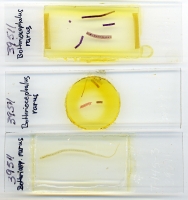
Fig. 29.-Section through cloacal region and cirrus sac.  -USNPC No. 39511 (types)
 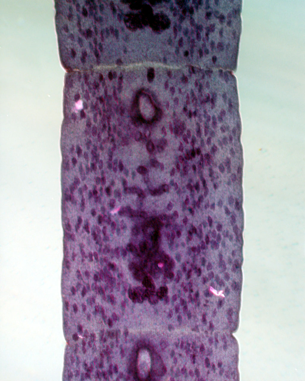 -USNPC No. 39511 (types)

