Line Drawing 1
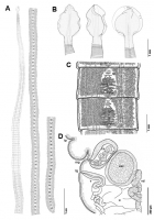
Line drawings of Adenocephalus pacificus ex Callorhinus ursinus from St. Paul Island, Alaska (AD) and holotype ex Arctocephalus philippii from Juan Fernández Islands, Chile (E). Whole worm, ventral v... MoreLine drawings of Adenocephalus pacificus ex Callorhinus ursinus from St. Paul Island, Alaska (AD) and holotype ex Arctocephalus philippii from Juan Fernández Islands, Chile (E). Whole worm, ventral view (A). Scoleces, lateral view (B). Mature segment, ventral view (D, E). Note: vitelline follicles omitted in right side of segment (D). |
Line Drawing 2
|
Photo Micrograph
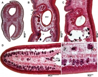
Photomicrographs of histological sections of Adenocephalus pacificus ex Callorhinus ursinus from St. Paul Island, Alaska. Cross-section showing of scolex shoving unicellular glands in the parenchyma o... MorePhotomicrographs of histological sections of Adenocephalus pacificus ex Callorhinus ursinus from St. Paul Island, Alaska. Cross-section showing of scolex shoving unicellular glands in the parenchyma of the scolex (A). Sagittal section through gravid proglottid (B, C). Cross-section of the gravid proglottid (D, E). Note: External seminal vesicle (esv); female gonopore (fg); numerous unicellular glands (g); male gonopore (mg); longitudinal musculature (lm); testes (t); transverse papilla-like tegumental protuberances (tp); uterus (u); vagina (v); vagina musculature (vm); viteline follicles (vf). |
Scanning Electron Micrograph
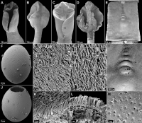
Scanning electron micrographs of Adenocephalus pacificus ex Callorhinus ursinus from St. Paul Island, Alaska. Scolex, dorsoventral view (A, C). Scolex, lateral view (B). Section of the scolex, lateral... MoreScanning electron micrographs of Adenocephalus pacificus ex Callorhinus ursinus from St. Paul Island, Alaska. Scolex, dorsoventral view (A, C). Scolex, lateral view (B). Section of the scolex, lateral view (D). Proglottid, ventral view (E). Detail of the genital pores with transverse papilla-like tegumental protuberance, ventral view (I). Egg (F, G). Detail of scolex surface converted with gladiate spinitriches, interspersed with much longer capilliform filitriches (G, H). Fraction of bothrial gladiate spinitriches (K). Fraction of the strobilar gladiate spinitriches (L). Detail of egg surface with pits (M). Note: small black letters correspond to the figures showing higher magnification images of these surfaces. Note: Female gonopore (fg); male gonopore (mg); transverse papilla-like tegumental protuberances (tp). |
 Line drawings of Adenocephalus pacificus ex Callorhinus ursinus from St. Paul Island, Alaska (AD) and holotype ex Arctocephalus philippii from Juan Fernández Islands, Chile (E). Whole worm, ventral view (A). Scoleces, lateral view (B). Mature segment, ventral view (D, E). Note: vitelline follicles omitted in right side of segment (D).
 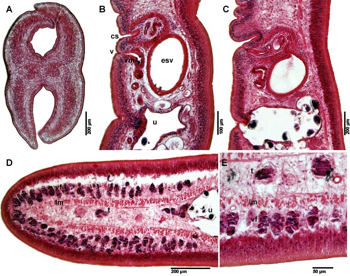 Photomicrographs of histological sections of Adenocephalus pacificus ex Callorhinus ursinus from St. Paul Island, Alaska. Cross-section showing of scolex shoving unicellular glands in the parenchyma of the scolex (A). Sagittal section through gravid proglottid (B, C). Cross-section of the gravid proglottid (D, E). Note: External seminal vesicle (esv); female gonopore (fg); numerous unicellular glands (g); male gonopore (mg); longitudinal musculature (lm); testes (t); transverse papilla-like tegumental protuberances (tp); uterus (u); vagina (v); vagina musculature (vm); viteline follicles (vf).
  Scanning electron micrographs of Adenocephalus pacificus ex Callorhinus ursinus from St. Paul Island, Alaska. Scolex, dorsoventral view (A, C). Scolex, lateral view (B). Section of the scolex, lateral view (D). Proglottid, ventral view (E). Detail of the genital pores with transverse papilla-like tegumental protuberance, ventral view (I). Egg (F, G). Detail of scolex surface converted with gladiate spinitriches, interspersed with much longer capilliform filitriches (G, H). Fraction of bothrial gladiate spinitriches (K). Fraction of the strobilar gladiate spinitriches (L). Detail of egg surface with pits (M). Note: small black letters correspond to the figures showing higher magnification images of these surfaces. Note: Female gonopore (fg); male gonopore (mg); transverse papilla-like tegumental protuberances (tp).

