Line Drawing 1

Figure 10. Paranoplocephala krebsi sp. nov. A, scolex. Byron Bay, host Dicrostonyx groenlandicus (scale bar=0.20 mm). B, genital ducts. Hope Bay, host D. groenlandicus (scale bar=0.20 mm). C, scolex. ... MoreFigure 10. Paranoplocephala krebsi sp. nov. A, scolex. Byron Bay, host Dicrostonyx groenlandicus (scale bar=0.20 mm). B, genital ducts. Hope Bay, host D. groenlandicus (scale bar=0.20 mm). C, scolex. Wrangel Island, host D. groenlandicus (scale bar=0.20 mm). D, early uterus in mature segment. Banks Island, host D. groenlandicus (scale bar=0.30 mm). E, egg and oncosphere with pyriform apparatus. Ungava Peninsula, host D. hudsonius (scale bar=0.040 mm). F, uterus in pregravid segment. Banks Island, host D. groenlandicus (scale bar=0.50 mm). |
Line Drawing 2
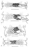
Figure 11. Paranoplocephala krebsi sp. nov.: mature segments. A, Byron Bay, host Dicrostonyx groenlandicus(scale bar=0.30 mm). B, Ungava Peninsula, host D. hudsonius (scale bar=0.30 mm). C, Wrangel Is... MoreFigure 11. Paranoplocephala krebsi sp. nov.: mature segments. A, Byron Bay, host Dicrostonyx groenlandicus(scale bar=0.30 mm). B, Ungava Peninsula, host D. hudsonius (scale bar=0.30 mm). C, Wrangel Island, host D.groenlandicus (scale bar=0.30 mm). D, Greenland, hostD. groenlandicus (scale bar=0.30 mm). |
Photo Micrograph
|
Scanning Electron Micrograph
|
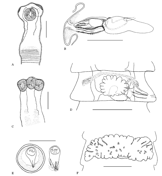 Figure 10. Paranoplocephala krebsi sp. nov. A, scolex. Byron Bay, host Dicrostonyx groenlandicus (scale bar=0.20 mm). B, genital ducts. Hope Bay, host D. groenlandicus (scale bar=0.20 mm). C, scolex. Wrangel Island, host D. groenlandicus (scale bar=0.20 mm). D, early uterus in mature segment. Banks Island, host D. groenlandicus (scale bar=0.30 mm). E, egg and oncosphere with pyriform apparatus. Ungava Peninsula, host D. hudsonius (scale bar=0.040 mm). F, uterus in pregravid segment. Banks Island, host D. groenlandicus (scale bar=0.50 mm).
  Figure 11. Paranoplocephala krebsi sp. nov.: mature segments. A, Byron Bay, host Dicrostonyx groenlandicus(scale bar=0.30 mm). B, Ungava Peninsula, host D. hudsonius (scale bar=0.30 mm). C, Wrangel Island, host D.groenlandicus (scale bar=0.30 mm). D, Greenland, hostD. groenlandicus (scale bar=0.30 mm).

