Line Drawing 1

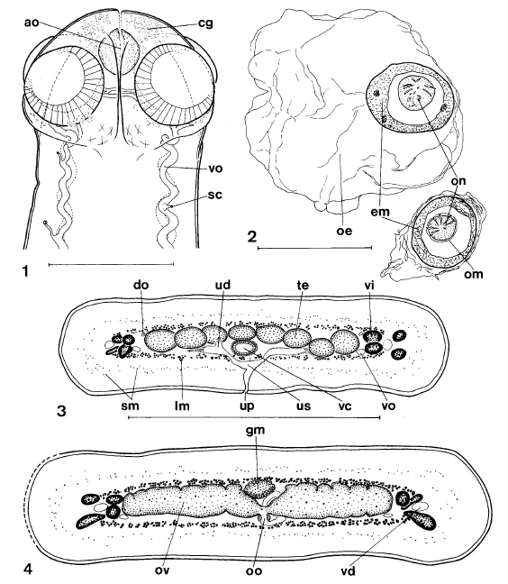
Figures 14. Nomimoscolex suspectus n. sp. 1. Scolex, ventral view, holotype, 34212 CHIOC. 2. Nomimoscolex suspectus n. sp. Eggs drawn in distilled water, paratype, 22302 INVE. 3. Nomimoscolex suspect... MoreFigures 14. Nomimoscolex suspectus n. sp. 1. Scolex, ventral view, holotype, 34212 CHIOC. 2. Nomimoscolex suspectus n. sp. Eggs drawn in distilled water, paratype, 22302 INVE. 3. Nomimoscolex suspectus n. sp. Transverse section of posterior part, mature proglottis, paratype, 27139 INVE. 4. Nomimoscolex suspectus n. sp. Transverse section at ovarian level, mature proglottis, paratype, 27139 INVE. Abbreviations: ao, apical organ; cg, glandular cells; do, dorsal osmoregulatory canal; em, embryophore; gm, Mehlis gland; lm, internal longitudinal musculature; oe, outer envelope; om, oncospheric membrane; on, oncosphere; oo, oöcapt; ov, ovary; sc, secondary canals; sm, secondary musculature; te, testes; ud, uterine diverticula; up, uterine pore; us, uterine stem; vc, vaginal canal; vd, vitelloduct; vi, vitelline follicles; vo, ventral osmoregulatory
canal: Scale-bars: 1, 250 µ; 2, 50 µm; 3,4, 500 µm. |
Line Drawing 2

Figures 56. Nomimoscolex suspectus n. sp. 5. Mature proglottis, dorsal view, from syntype material of N. piraeeba Woodland (1934, plate 10, figure 7), BMNH 1964.12.15.111-122. Abbreviations: vs, vagi... MoreFigures 56. Nomimoscolex suspectus n. sp. 5. Mature proglottis, dorsal view, from syntype material of N. piraeeba Woodland (1934, plate 10, figure 7), BMNH 1964.12.15.111-122. Abbreviations: vs, vaginal sphincter. 6. Nomimoscolex suspectus n. sp. Holotype, 34212 CHIOC, dorsal view, mature proglottis. Scale-bar: 500 µm. |
Photo Micrograph
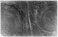
Figure 7. Nomimoscolex suspectus n. sp. Photomicrograph of scolex. Detail of suckers showing the numerous spiniform microtriches on their rim and the smaller spiniform microtriches present on the scol... MoreFigure 7. Nomimoscolex suspectus n. sp. Photomicrograph of scolex. Detail of suckers showing the numerous spiniform microtriches on their rim and the smaller spiniform microtriches present on the scolex. The spiniform microtriches end at the level of the posterior margin of the sucker (arrow). Holotype 34212 CHIOC. Scale-bar: 50 µm. |
Scanning Electron Micrograph
|
 Figures 14. Nomimoscolex suspectus n. sp. 1. Scolex, ventral view, holotype, 34212 CHIOC. 2. Nomimoscolex suspectus n. sp. Eggs drawn in distilled water, paratype, 22302 INVE. 3. Nomimoscolex suspectus n. sp. Transverse section of posterior part, mature proglottis, paratype, 27139 INVE. 4. Nomimoscolex suspectus n. sp. Transverse section at ovarian level, mature proglottis, paratype, 27139 INVE. Abbreviations: ao, apical organ; cg, glandular cells; do, dorsal osmoregulatory canal; em, embryophore; gm, Mehlis gland; lm, internal longitudinal musculature; oe, outer envelope; om, oncospheric membrane; on, oncosphere; oo, oöcapt; ov, ovary; sc, secondary canals; sm, secondary musculature; te, testes; ud, uterine diverticula; up, uterine pore; us, uterine stem; vc, vaginal canal; vd, vitelloduct; vi, vitelline follicles; vo, ventral osmoregulatory
canal: Scale-bars: 1, 250 µ; 2, 50 µm; 3,4, 500 µm.
Figures 14. Nomimoscolex suspectus n. sp. 1. Scolex, ventral view, holotype, 34212 CHIOC. 2. Nomimoscolex suspectus n. sp. Eggs drawn in distilled water, paratype, 22302 INVE. 3. Nomimoscolex suspectus n. sp. Transverse section of posterior part, mature proglottis, paratype, 27139 INVE. 4. Nomimoscolex suspectus n. sp. Transverse section at ovarian level, mature proglottis, paratype, 27139 INVE. Abbreviations: ao, apical organ; cg, glandular cells; do, dorsal osmoregulatory canal; em, embryophore; gm, Mehlis gland; lm, internal longitudinal musculature; oe, outer envelope; om, oncospheric membrane; on, oncosphere; oo, oöcapt; ov, ovary; sc, secondary canals; sm, secondary musculature; te, testes; ud, uterine diverticula; up, uterine pore; us, uterine stem; vc, vaginal canal; vd, vitelloduct; vi, vitelline follicles; vo, ventral osmoregulatory
canal: Scale-bars: 1, 250 µ; 2, 50 µm; 3,4, 500 µm. 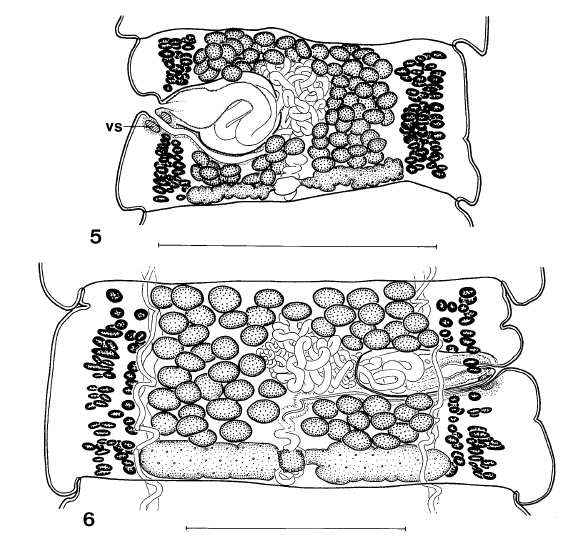 Figures 56. Nomimoscolex suspectus n. sp. 5. Mature proglottis, dorsal view, from syntype material of N. piraeeba Woodland (1934, plate 10, figure 7), BMNH 1964.12.15.111-122. Abbreviations: vs, vaginal sphincter. 6. Nomimoscolex suspectus n. sp. Holotype, 34212 CHIOC, dorsal view, mature proglottis. Scale-bar: 500 µm.
 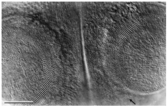 Figure 7. Nomimoscolex suspectus n. sp. Photomicrograph of scolex. Detail of suckers showing the numerous spiniform microtriches on their rim and the smaller spiniform microtriches present on the scolex. The spiniform microtriches end at the level of the posterior margin of the sucker (arrow). Holotype 34212 CHIOC. Scale-bar: 50 µm.

