Line Drawing 1
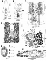
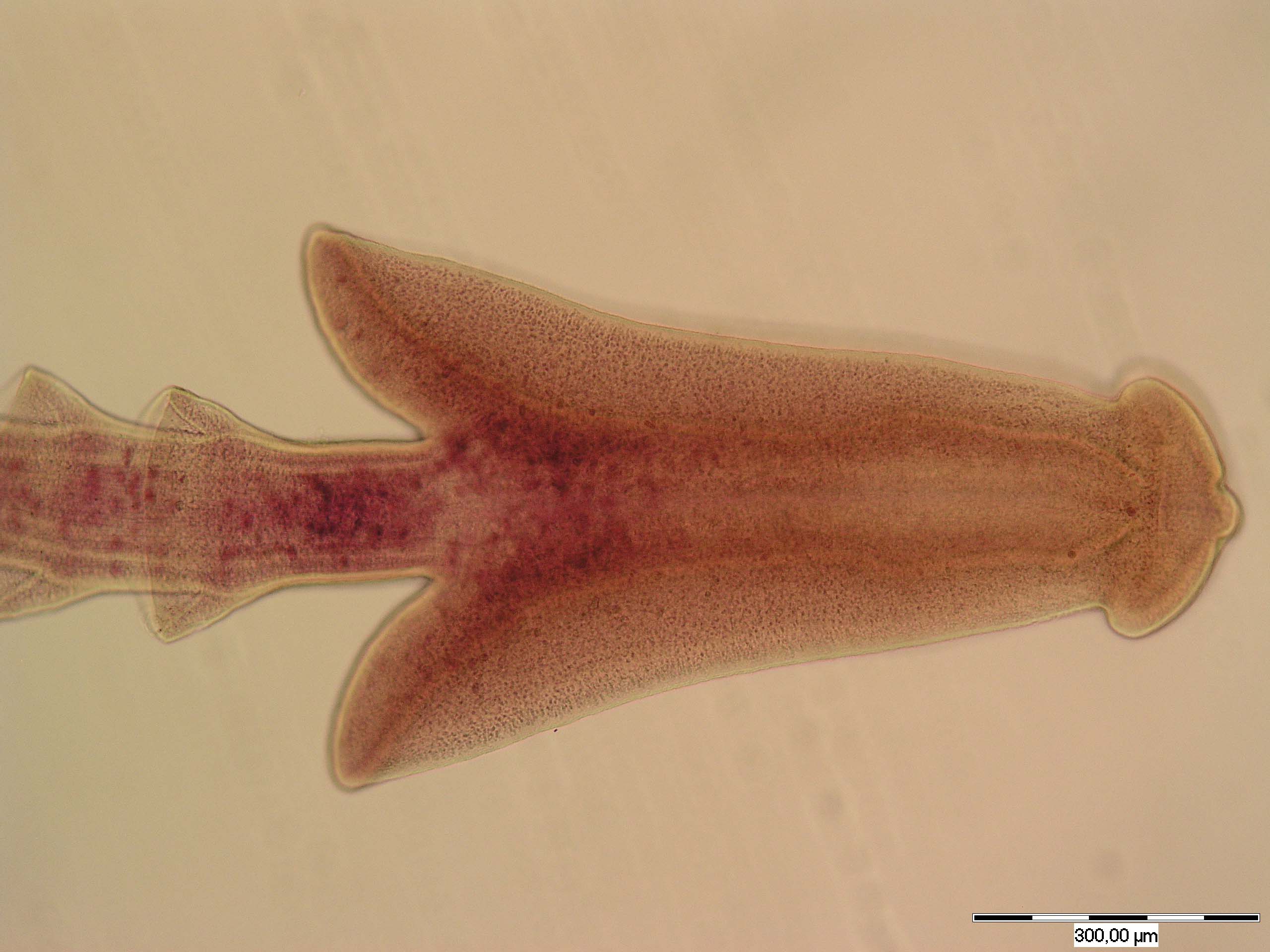
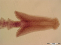
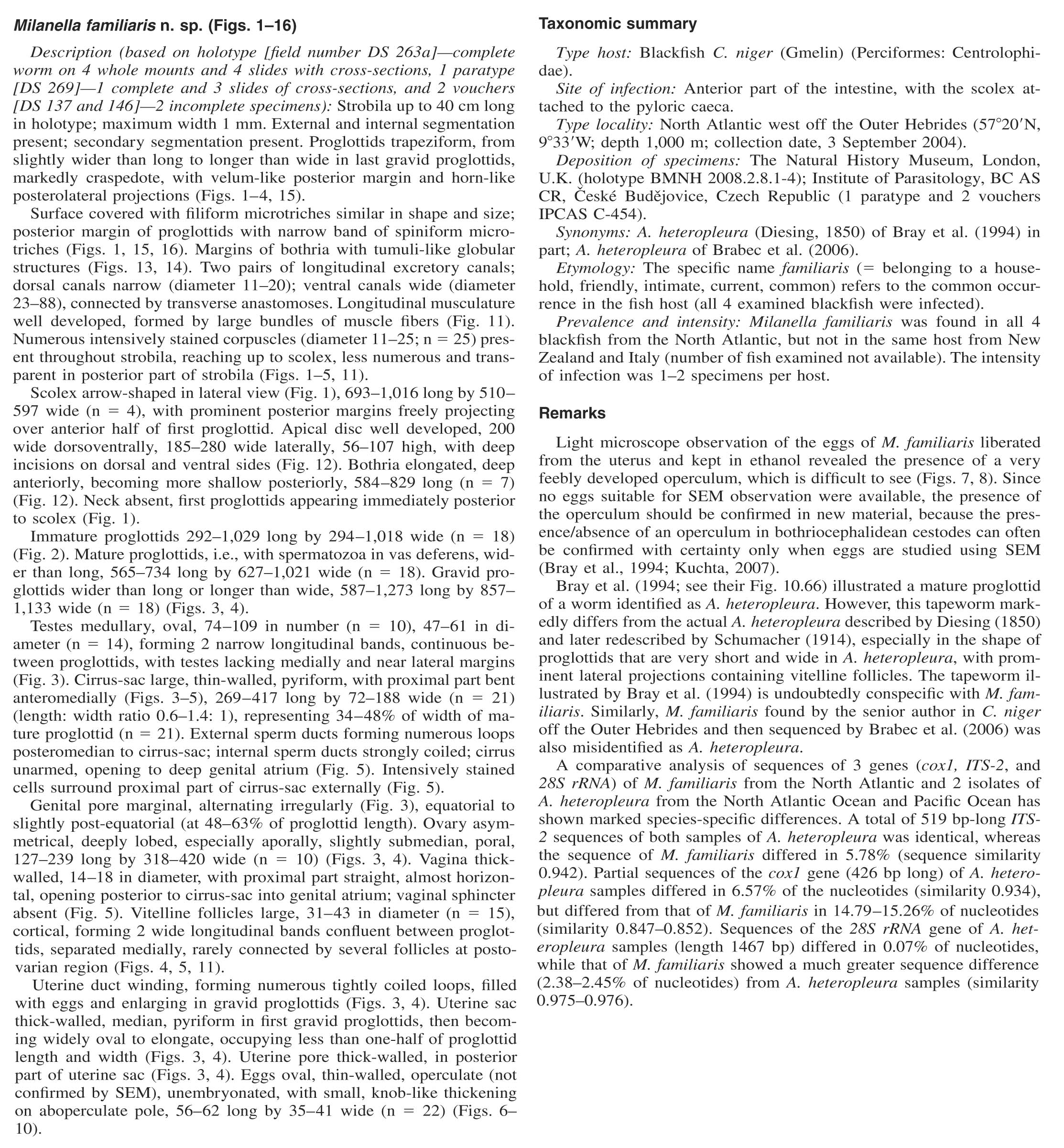
FIGURES 111. Milanella familiaris n. gen., n. sp., holotype (BMNH 2008.2.8.1-4). (1) Scolex of holotype with intensively stained corpuscles;
lateral view. Insetspiniform microtriches on the posteri... MoreFIGURES 111. Milanella familiaris n. gen., n. sp., holotype (BMNH 2008.2.8.1-4). (1) Scolex of holotype with intensively stained corpuscles;
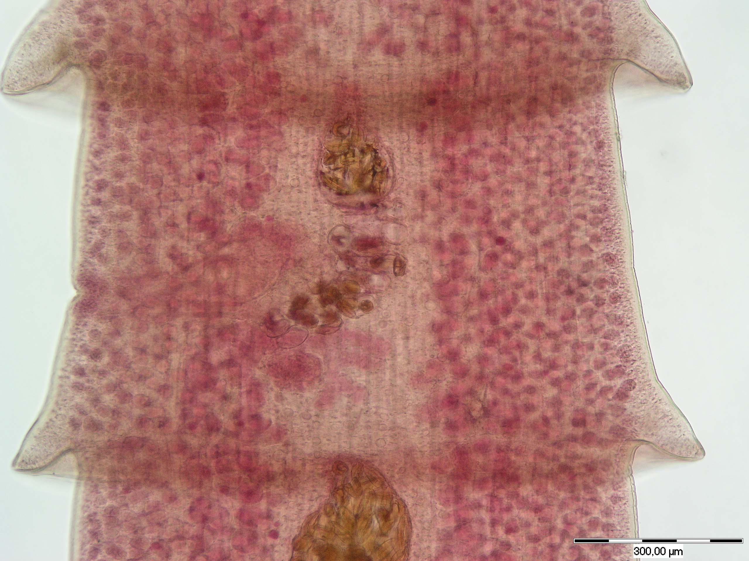
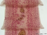
lateral view. Insetspiniform microtriches on the posterior margins of proglottids. (2) Anterior part of strobila with intensively stained corpuscles. First proglottiddorsal, secondventral, and thirdmedian view. (3) First gravid proglottids. Ventral view with intensively stained corpuscles.
In second segment, vitelline follicles not illustrated. (4) Schematic drawing of gravid proglottid. Ventral view; testes not illustrated. (5) Terminal
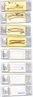
genitalia. Ventral view. (610) Freshly liberated eggs, fixed by ethanol. (6) Whole egg. (7, 8) Details of operculum. (9, 10) Details of knob-like
thickening on aboperculate pole. (11) Cross-section at the level of cirrus-sac. Abbreviations: c, cirrus; co, corpuscles; cs, cirrus-sac; doc, dorsal
osmoregulatory canal; e, eggs; ga, genital atrium; ilm, inner longitudinal muscles; ov, ovary; t, testes; up, uterine pore; us, uterine sac; va, vagina; vf, vitelline follicles; voc, ventral osmoregulatory canal. |
Line Drawing 2
|
Photo Micrograph

FIGURES 1216. Scanning electron micrographs of M. familiaris n. gen., n. sp. (12) Scolex; dorsoventral view. Number indicates where Figure
13 was taken. (13) Distal surface of bothria. Number indica... MoreFIGURES 1216. Scanning electron micrographs of M. familiaris n. gen., n. sp. (12) Scolex; dorsoventral view. Number indicates where Figure
13 was taken. (13) Distal surface of bothria. Number indicates where Figure 14 was taken. (14) Detail of tumuli-like, globular structures. (15)
Gravid proglottids; dorsal view. Number indicates where Figure 16 was taken. (16) Detailed view of posterior margin of proglottid. |
Scanning Electron Micrograph
|

 FIGURES 111. Milanella familiaris n. gen., n. sp., holotype (BMNH 2008.2.8.1-4). (1) Scolex of holotype with intensively stained corpuscles;
lateral view. Insetspiniform microtriches on the posterior margins of proglottids. (2) Anterior part of strobila with intensively stained corpuscles. First proglottiddorsal, secondventral, and thirdmedian view. (3) First gravid proglottids. Ventral view with intensively stained corpuscles.
In second segment, vitelline follicles not illustrated. (4) Schematic drawing of gravid proglottid. Ventral view; testes not illustrated. (5) Terminal
genitalia. Ventral view. (610) Freshly liberated eggs, fixed by ethanol. (6) Whole egg. (7, 8) Details of operculum. (9, 10) Details of knob-like
thickening on aboperculate pole. (11) Cross-section at the level of cirrus-sac. Abbreviations: c, cirrus; co, corpuscles; cs, cirrus-sac; doc, dorsal
osmoregulatory canal; e, eggs; ga, genital atrium; ilm, inner longitudinal muscles; ov, ovary; t, testes; up, uterine pore; us, uterine sac; va, vagina; vf, vitelline follicles; voc, ventral osmoregulatory canal.
FIGURES 111. Milanella familiaris n. gen., n. sp., holotype (BMNH 2008.2.8.1-4). (1) Scolex of holotype with intensively stained corpuscles;
lateral view. Insetspiniform microtriches on the posterior margins of proglottids. (2) Anterior part of strobila with intensively stained corpuscles. First proglottiddorsal, secondventral, and thirdmedian view. (3) First gravid proglottids. Ventral view with intensively stained corpuscles.
In second segment, vitelline follicles not illustrated. (4) Schematic drawing of gravid proglottid. Ventral view; testes not illustrated. (5) Terminal
genitalia. Ventral view. (610) Freshly liberated eggs, fixed by ethanol. (6) Whole egg. (7, 8) Details of operculum. (9, 10) Details of knob-like
thickening on aboperculate pole. (11) Cross-section at the level of cirrus-sac. Abbreviations: c, cirrus; co, corpuscles; cs, cirrus-sac; doc, dorsal
osmoregulatory canal; e, eggs; ga, genital atrium; ilm, inner longitudinal muscles; ov, ovary; t, testes; up, uterine pore; us, uterine sac; va, vagina; vf, vitelline follicles; voc, ventral osmoregulatory canal. 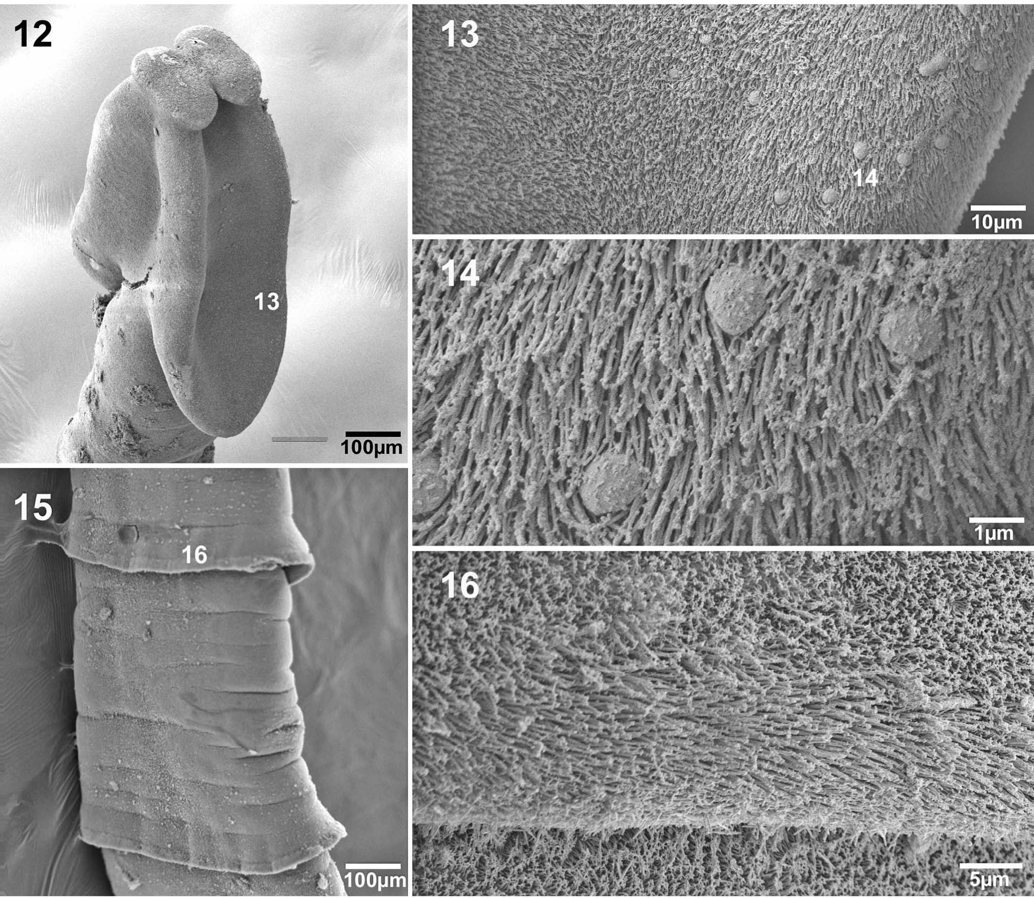 FIGURES 1216. Scanning electron micrographs of M. familiaris n. gen., n. sp. (12) Scolex; dorsoventral view. Number indicates where Figure
13 was taken. (13) Distal surface of bothria. Number indicates where Figure 14 was taken. (14) Detail of tumuli-like, globular structures. (15)
Gravid proglottids; dorsal view. Number indicates where Figure 16 was taken. (16) Detailed view of posterior margin of proglottid.
FIGURES 1216. Scanning electron micrographs of M. familiaris n. gen., n. sp. (12) Scolex; dorsoventral view. Number indicates where Figure
13 was taken. (13) Distal surface of bothria. Number indicates where Figure 14 was taken. (14) Detail of tumuli-like, globular structures. (15)
Gravid proglottids; dorsal view. Number indicates where Figure 16 was taken. (16) Detailed view of posterior margin of proglottid. 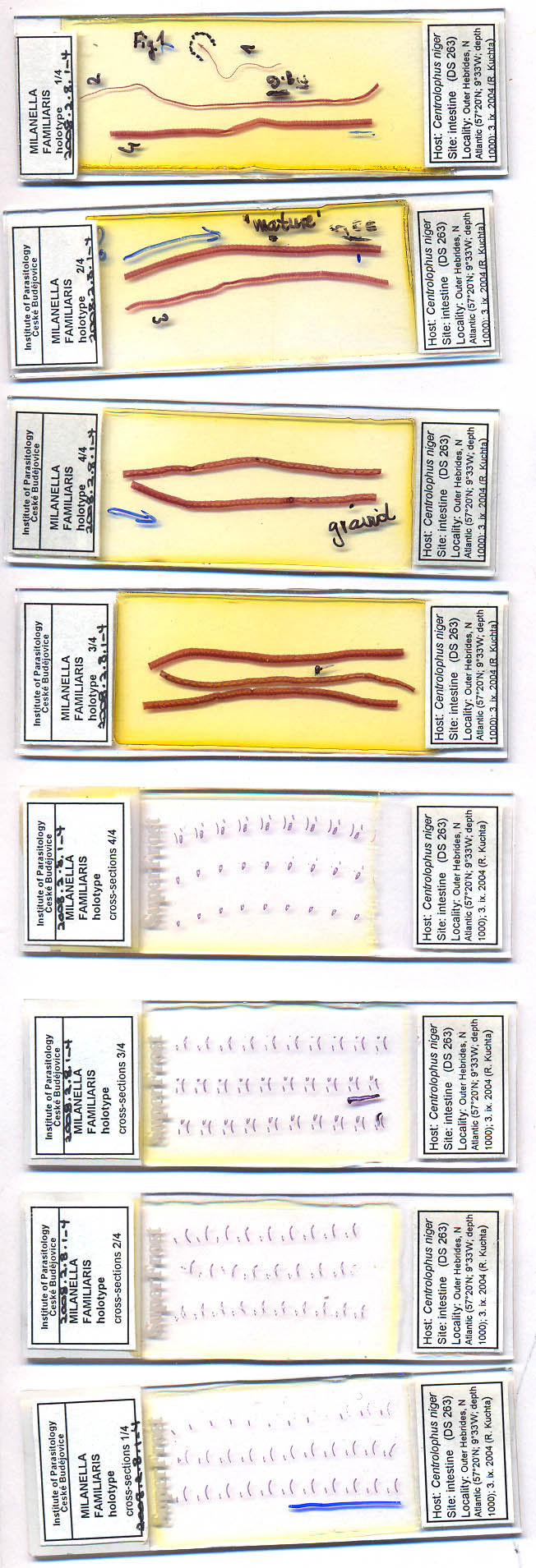 BMNH 2008.2.8.1-4