Cestode Scientific Name
| Species ID | 5837 |
|---|---|
| Order | Bothriocephalidea |
| Family | Bothriocephalidae |
| Subfamily | |
| Genus | Anantrum |
| Species | tortum |
| Authority | (Linton, 1905) Overstreet, 1968 |
| Taxonomic Status | Valid |
| Valid Name | |
| Synonyms | Dibothrium tortum Linton, 1905 |
| Genus Record | No |
| Type Species | Yes |
| Verified | Yes |
| Verified By | R. Kuchta |
| Citation(s) |
Linton, E. 1905. Parasites of fishes of Beaufort, North Carolina. Bulletin of the United States Bureau of Fisheries (1904) 24: 321-428. (275) Download PDFOverstreet, R. M. 1968. Parasites of the inshore lizardfish, Synodus foetens, from south Florida, including a description of a new genus of cestoda. Bulletin of Marine Science 18(2): 444-470. (4557) Download PDF |
| Redescription |
Rees, G. 1969. Cestodes from Bermuda fishes and an account of Acompsocephalum tortum (Linton, 1905) gen. nov. from the lizard fish Synodus intermedius (Agassiz). Parasitology 59: 519-548. (369) Download PDF |
| Scientific Name Notes |
Record Data
| Date (MM/DD/YYYY) | Action | User Name |
|---|---|---|
| 03/05/2010 | Created | B. Barbeau |
| 06/24/2014 | Modified | |
| 01/25/2016 | Modified | K. Mojica |
| 01/28/2016 | Modified | K. Mojica |
| 09/01/2017 | Modified | R. Kuchta |
| 09/18/2017 | Modified | R. Kuchta |
| 05/16/2024 | Modified | R. Kuchta |
Type Host
| Host Class | Actinopterygii | ||||||
|---|---|---|---|---|---|---|---|
| Host Order | Aulopiformes | ||||||
| Host Family | Synodontidae | ||||||
|
Type Host (Literal) |
|
||||||
|
Type Host (Valid) |
|
||||||
| Additional Host(s) | |||||||
| Site in Host | intestine | ||||||
| Host Notes |
Type Locality
| Country | U.S.A. |
|---|---|
| Body of Water | Atlantic Ocean |
| Island(s) | |
| City/Region | Buttinwood Canal, North Carolina |
| Coordinates | |
| DD Latitude | |
| DD Longitude | |
| Additional Localities | |
| Locality Notes |
Specimens
| Type Material | -USNPC No. 71122 (type) |
|---|---|
| Total Number of Type Specimens | |
| Voucher Material | |
| Specimen Notes |
Data are given as in original description unless otherwise indicated.
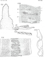
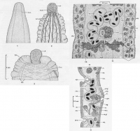
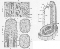
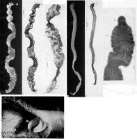

 Fig. 119. Sketch from life enlarged. Most specimens are relatively more slender than the one represented in the sketch. Anterior egg clusters nearly transverse, succeeding ones becoming diagonal.
Fig. 120. Fragment from posterior end with egg clusters but little inclined to the axes of the body.
Fig. 121. Anterior end of living specimen. Actual diameter at r, 0.45 mm.
Fig. 122. Anterior end of alcoholic specimen. Actual diameter at anterior end 0.36 mm.
Fig. 123. Sketch of body with three sets of genitalia. Specimen mounted in balsam. Actual breadth 1.77 mm.
Fig. 124. Sketch of two sets of genitalia. Life, with few details added from stained specimens x 300.
Fig. 119. Sketch from life enlarged. Most specimens are relatively more slender than the one represented in the sketch. Anterior egg clusters nearly transverse, succeeding ones becoming diagonal.
Fig. 120. Fragment from posterior end with egg clusters but little inclined to the axes of the body.
Fig. 121. Anterior end of living specimen. Actual diameter at r, 0.45 mm.
Fig. 122. Anterior end of alcoholic specimen. Actual diameter at anterior end 0.36 mm.
Fig. 123. Sketch of body with three sets of genitalia. Specimen mounted in balsam. Actual breadth 1.77 mm.
Fig. 124. Sketch of two sets of genitalia. Life, with few details added from stained specimens x 300. 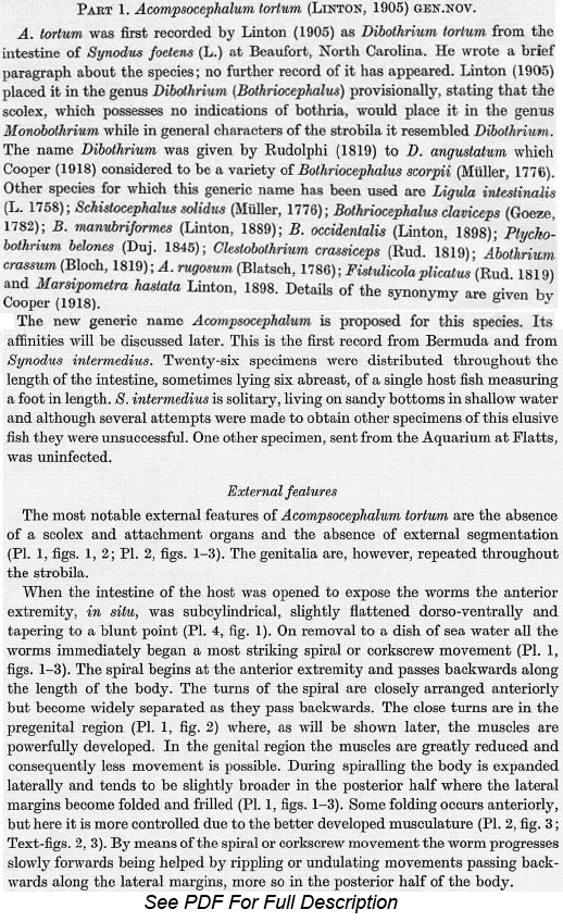
 Text-fig. 1. Anterior extremity, 'resting state'.
Text-fig. 2. Anterior extremity, contracted, showing 'papilla', folded margins of strobila and osmoregulatory system. Text-fig. 3. Anterior extremity, contracted still further, showing greater folding of the lateral margins and the osmoregulatory canals. Text-fig. 4. Acompscocephalum tortum (Linton). One set of mature reproductive organs, dorsal view. Text-fig. 5. Sagittal section showing reproductive system. The two interruptions indicate that the ventral uterine pore is not in line with the dorsal genital atrium.
Text-fig. 1. Anterior extremity, 'resting state'.
Text-fig. 2. Anterior extremity, contracted, showing 'papilla', folded margins of strobila and osmoregulatory system. Text-fig. 3. Anterior extremity, contracted still further, showing greater folding of the lateral margins and the osmoregulatory canals. Text-fig. 4. Acompscocephalum tortum (Linton). One set of mature reproductive organs, dorsal view. Text-fig. 5. Sagittal section showing reproductive system. The two interruptions indicate that the ventral uterine pore is not in line with the dorsal genital atrium.  Test-fig. 6. Early genital region showing osmoregulatory canals, two sets of developing reproductive organs and absence of segmentation. Text-fig. 7. Transverse section near anterior extremity showing lateral cerebral ganglion, ring commissures and origin of accessory lateral nerves. Text-fig. 8. Transverse section close behind the previous showing transverse commissure and network of the osmoregulatory system. Text-fig. 9. Sagittal section showing fanning of longitudinal muscles, part of the two accessory lateral nerves of one side and main lateral nerve cord, osmoregulatory system and apical processes. Text-fig. 10. Reconstruction of nervous system at anterior extremity and longitudinal muscles forming a ring before they fan out towards the surface.
  Plate 1. Fig. 1. Preserved specimen showing spiral movements, spiral more marked anteriorly, frilled margins more marked posteriorly. Anterior papilla and absence of segmentation. Fig. 2. Same specimen as previous, stained and mounted, showing repetition of reproductive organs, pregenital, early genital and mature regions.
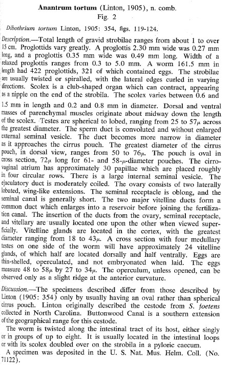
Best viewed in Firefox


