Line Drawing 1
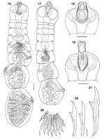
Figs. 1622 Paraprogynotaenia minuta n. sp. from Tringa sp. in France (except Fig. 17, which shows the holotype from Charadrius alexandrinus in Bulgaria). 16, 17. Entire individuals, dorsal view. ... MoreFigs. 1622 Paraprogynotaenia minuta n. sp. from Tringa sp. in France (except Fig. 17, which shows the holotype from Charadrius alexandrinus in Bulgaria). 16, 17. Entire individuals, dorsal view. 18. Scolex with retracted rostellum (rostellar hooks partially lost), optical section. 19. Scolex with protracted rostellum (rostellar hooks lost), optical section. 20. Semicircle of seven rostellar hooks with two lateral hooks on each side. 21. Rostellar hook from a semicircle. 22. Lateral rostellar hooks. Scale-bars: 1619, 100 µm; 20, 20 µm; 21,22, 10 µm. |
Line Drawing 2

Figs. 2327 Paraprogynotaenia minuta n. sp. from Tringa sp. in France. 23. Female and post-mature proglottides, dorsal view. 24. Female proglottis (detail), dorsal view. Abbreviations: Ot, oötype;... MoreFigs. 2327 Paraprogynotaenia minuta n. sp. from Tringa sp. in France. 23. Female and post-mature proglottides, dorsal view. 24. Female proglottis (detail), dorsal view. Abbreviations: Ot, oötype; Ov, ovary; SR, seminal receptacle; SRd, duct of seminal
receptacle; Ut, uterus; Utd, uterine duct; Vt, vitellarium. 25. Male proglottis, dorsally. 26. Gravid proglottis, dorsally. 27. Eggs. Scalebars:
23, 50 µm; 24, 20 µm; 25,26, 100 µm; 27, 10 µm |
Photo Micrograph
|
Scanning Electron Micrograph
|
 Figs. 1622 Paraprogynotaenia minuta n. sp. from Tringa sp. in France (except Fig. 17, which shows the holotype from Charadrius alexandrinus in Bulgaria). 16, 17. Entire individuals, dorsal view. 18. Scolex with retracted rostellum (rostellar hooks partially lost), optical section. 19. Scolex with protracted rostellum (rostellar hooks lost), optical section. 20. Semicircle of seven rostellar hooks with two lateral hooks on each side. 21. Rostellar hook from a semicircle. 22. Lateral rostellar hooks. Scale-bars: 1619, 100 µm; 20, 20 µm; 21,22, 10 µm.
 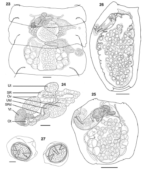 Figs. 2327 Paraprogynotaenia minuta n. sp. from Tringa sp. in France. 23. Female and post-mature proglottides, dorsal view. 24. Female proglottis (detail), dorsal view. Abbreviations: Ot, oötype; Ov, ovary; SR, seminal receptacle; SRd, duct of seminal
receptacle; Ut, uterus; Utd, uterine duct; Vt, vitellarium. 25. Male proglottis, dorsally. 26. Gravid proglottis, dorsally. 27. Eggs. Scalebars:
23, 50 µm; 24, 20 µm; 25,26, 100 µm; 27, 10 µm
Figs. 2327 Paraprogynotaenia minuta n. sp. from Tringa sp. in France. 23. Female and post-mature proglottides, dorsal view. 24. Female proglottis (detail), dorsal view. Abbreviations: Ot, oötype; Ov, ovary; SR, seminal receptacle; SRd, duct of seminal
receptacle; Ut, uterus; Utd, uterine duct; Vt, vitellarium. 25. Male proglottis, dorsally. 26. Gravid proglottis, dorsally. 27. Eggs. Scalebars:
23, 50 µm; 24, 20 µm; 25,26, 100 µm; 27, 10 µm 

