Line Drawing 1

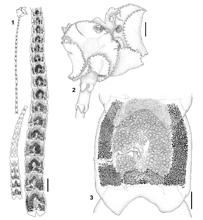
FIGURES 13. Crossobothrium antonioi n. sp. (1) Entire worm, scale bar = 1.6 mm. (2) Scolex, scale bar = 200 µm. (3) Mature proglottid, scale bar = 400 µm. |
Line Drawing 2
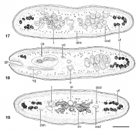
FIGURES 1719. Crossobothrium antonioi n. sp., cross sections of mature proglottid, scale bar = 200 µm. (17) At level of testes anterior to cirrus sac. (18) At level of genital pore. (19) At level of ... MoreFIGURES 1719. Crossobothrium antonioi n. sp., cross sections of mature proglottid, scale bar = 200 µm. (17) At level of testes anterior to cirrus sac. (18) At level of genital pore. (19) At level of ovarian isthmus. Abbreviations: cs, cirrus sac; dlm, deep longitudinal musculature; dod, dorsal osmoregulatory duct; gp, genital pore; ov, ovary; sr, seminal receptacle; t, testis; u, uterus; vd, vas deferens; vf, vitelline follicle; vg, vagina; vod, ventral osmoregulatory duct. |
Photo Micrograph
|
Scanning Electron Micrograph
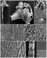
FIGURES 416. Crossobothrium antonioi n. sp., scanning electron micrographs. (4) General view of scolex and immature proglottids, scale bar = 500 µm. (5) Scolex, scale bar = 200 µm. Small numbers indi... MoreFIGURES 416. Crossobothrium antonioi n. sp., scanning electron micrographs. (4) General view of scolex and immature proglottids, scale bar = 500 µm. (5) Scolex, scale bar = 200 µm. Small numbers indicate locations of details shown in Figures 913. (6) Detail of accessory sucker, scale bar = 50 µm. Small numbers indicate locations of details shown in Figures 7 and 8. (7) Inner surface of accessory sucker, scale bar = 1 µm. (8) Outer surface of accessory sucker, scale bar = 1 µm. (9) Distal bothridial surface showing gradual change on microthrix size and density, sale bar = 10 µm. (10) Distal bothridial surface near the margin, scale bar = 2 µm. (11) Distal bothridial surface at midway between margin and center, scale bar = 2 µm. (12) Distal bothridial surface at the center of bothridium, scale bar = 2 µm. (13) Proximal bothridial surface (a), scale bar = 2 µm; detail of microtriches in close up (b), scale bar = 1 µm. (14) Surface of immature proglottids, scale bar = 200 µm. Small numbers indicate locations of details shown in Figures 15 and 16. (15) Surface of laciniations in immature proglottid, scale bar = 2 µm. (16) Surface of immature proglottid, scale bar = 2 µm. |

 FIGURES 13. Crossobothrium antonioi n. sp. (1) Entire worm, scale bar = 1.6 mm. (2) Scolex, scale bar = 200 µm. (3) Mature proglottid, scale bar = 400 µm.
  FIGURES 1719. Crossobothrium antonioi n. sp., cross sections of mature proglottid, scale bar = 200 µm. (17) At level of testes anterior to cirrus sac. (18) At level of genital pore. (19) At level of ovarian isthmus. Abbreviations: cs, cirrus sac; dlm, deep longitudinal musculature; dod, dorsal osmoregulatory duct; gp, genital pore; ov, ovary; sr, seminal receptacle; t, testis; u, uterus; vd, vas deferens; vf, vitelline follicle; vg, vagina; vod, ventral osmoregulatory duct.
 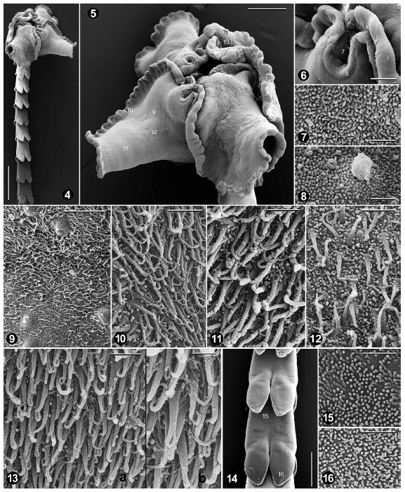 FIGURES 416. Crossobothrium antonioi n. sp., scanning electron micrographs. (4) General view of scolex and immature proglottids, scale bar = 500 µm. (5) Scolex, scale bar = 200 µm. Small numbers indicate locations of details shown in Figures 913. (6) Detail of accessory sucker, scale bar = 50 µm. Small numbers indicate locations of details shown in Figures 7 and 8. (7) Inner surface of accessory sucker, scale bar = 1 µm. (8) Outer surface of accessory sucker, scale bar = 1 µm. (9) Distal bothridial surface showing gradual change on microthrix size and density, sale bar = 10 µm. (10) Distal bothridial surface near the margin, scale bar = 2 µm. (11) Distal bothridial surface at midway between margin and center, scale bar = 2 µm. (12) Distal bothridial surface at the center of bothridium, scale bar = 2 µm. (13) Proximal bothridial surface (a), scale bar = 2 µm; detail of microtriches in close up (b), scale bar = 1 µm. (14) Surface of immature proglottids, scale bar = 200 µm. Small numbers indicate locations of details shown in Figures 15 and 16. (15) Surface of laciniations in immature proglottid, scale bar = 2 µm. (16) Surface of immature proglottid, scale bar = 2 µm.

