Cestode Scientific Name
| Species ID | 5791 |
|---|---|
| Order | Onchoproteocephalidea II |
| Family | |
| Subfamily | |
| Genus | Acanthobothrium |
| Species | rodmani |
| Authority | Fyler, Caira & Jensen, 2009 |
| Taxonomic Status | Valid |
| Valid Name | |
| Synonyms | |
| Genus Record | No |
| Type Species | No |
| Verified | No |
| Verified By | |
| Citation(s) |
Fyler, C. A., J. N. Caira, and K. Jensen. 2009. Five new species of Acanthobothrium (Cestoda: Tetraphyllidea) from an unusual species of Himantura (Rajiformes: Dasyatidae) from northern Australia. Folia Parasitologica 56(2): 107-128. (4538) Download PDF |
| Redescription | |
| Scientific Name Notes |
Record Data
| Date (MM/DD/YYYY) | Action | User Name |
|---|---|---|
| 12/21/2009 | Created | B. Barbeau |
| 02/09/2010 | Modified | |
| 06/07/2015 | Modified | T. Ruhnke |
| 01/22/2016 | Modified | K. Mojica |
| 09/01/2016 | Modified | K. Jensen |
Type Host
| Host Class | |||||||
|---|---|---|---|---|---|---|---|
| Host Order | Myliobatiformes | ||||||
| Host Family | Dasyatidae | ||||||
|
Type Host (Literal) |
|
||||||
|
Type Host (Valid) |
|
||||||
| Additional Host(s) | |||||||
| Site in Host | spiral intestine | ||||||
| Host Notes |
Type Locality
| Country | Australia |
|---|---|
| Body of Water | Arafura Sea, East of Wessel Islands |
| Island(s) | |
| City/Region | Northern Territory |
| Coordinates | |
| DD Latitude | -11.296 |
| DD Longitude | 136.997 |
| Additional Localities | |
| Locality Notes |
Specimens
| Type Material | QM No. G231354 (holotype); QM Nos. G231355-G231357 (3 paratypes); USNPC Nos. 101961-101963 (3 paratypes); LRP Nos. 4333-4335 (3 paratypes); QM Nos. G231359 (cross-sections of proglottid of 1 paratype and corresponding voucher QM G231358); LRP Nos. 4564-4569 (cross-sections of scolex of 1 paratype and corresponding voucehr LRP 4563); LRP Nos. 4560-4562 (longitudinal sections of scolex of 1 paratype and corresponding voucher LRP 4559); LRP Nos. 4336-4339 (3 paratypes prepared for SEM); LRP No. 4340: GenBank No. FJ843596 [CF-87], LRP No. 4341: GenBank No. FJ843597 [CF-124] (2 paratypes of hologenophores). |
|---|---|
| Total Number of Type Specimens | whole mounts of 10 mature worms, cross-sections of mature proglottids of 1 worm and whole mount of its voucher, 4 scoleces examined with SEM and whole mount of their vouchers, cross-sections of scolex of 1 worm and whole mount of its voucher, longitudinal sections of scolex of 1 worm and whole mount of its voucher, and the scoleces and posterior portion of the strobila of 2 hologenephores. |
| Voucher Material | IPCAS No. C-524 (2 vouchers). |
| Specimen Notes |
Data are given as in original description unless otherwise indicated.
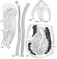
 Figs. 19-22. Line drawings of Acanthobothrium rodmani sp. n. Fig. 19. Scolex (QM G231354). Arrowheads indicate location of cross-sections in Figs. 52 and 53. Fig. 20. Hooks (QM G231354). Fig. 21. Terminal proglotiids (LRP 4333). Arrowhead indicates location of cross-section shown in Fig. 51. Fig. 22. Whole worm (QM G231354).
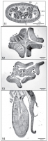
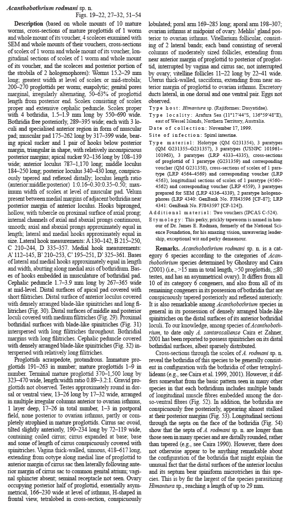
  Fig. 51-54. Histological sections. Fig. 51. Cross-section through ovary of Acanthobothrium rodmani sp. n. (QM G231359). Fig. 52. Cross-section through anterior region of anterior loculus of A. rodmani sp. n. (LRP 4564). Fig. 53. Cross-section through posterior region of anterior loculus of A. rodmani sp. n. (LRP 4566). Arrowheads indicate posterior stalk-like connection between bothridium and cephalic peduncle. Fig. 54. Longitudinal section through scolex showing bothridial septa of A. rodmani sp. n. (LRP 4561). Arrowheads indicate location of septa dividing adjacent bothridial loculi. Abbreviations: DED - dorsal excretory duct; MG - Mehlis´ gland; O - ovary; U - uterus; V - vitelline follicle; VED - ventral excretory duct.
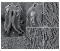
 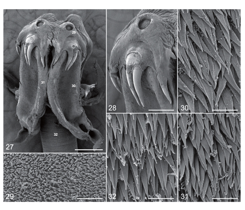 Figs. 27-32. Scanning electron micrographs of Acanthobothrium rodmani sp. n. Fig. 27. Scolex. Note: small numbers correspond to the figures showing higher magnification images of these surfaces. Fig. 28. Detail of apical pad and hooks. Fig. 29. Surface of apical pad. Fig. 30. Distal bothridial surface. Fig. 31. Proximal bothridial surface. Fig. 32. Cephalic peduncle surface. Scale bars: Fig. 27 = 200µm; Fig. 28 = 100µm; Fig. 29-32 = 2µm.
 Best viewed in Firefox


