Line Drawing 1
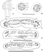
Fig. 1 Luciaella ivanovae n. g., n. sp. from Ageneiosus inermis. A, scolex in dorsoventral view showing gland cells; B, spontaneously laid eggs; C, transverse section of mature proglottis at level of ... MoreFig. 1 Luciaella ivanovae n. g., n. sp. from Ageneiosus inermis. A, scolex in dorsoventral view showing gland cells; B, spontaneously laid eggs; C, transverse section of mature proglottis at level of ovary; D,E, transverse sections of last immature proglottis and gravid proglottides at level of testes, showing the development of the uterus. Abbreviations: doc, dorsal osmoregulatory canal; em, embryophore; gc, gland cells; lh, larval hooks; lm, longitudinal musculature; oe, outer envelope; on, oncosphere; ov, ovary; te, testis; ut, uterus; vf, vitelline follicles; voc, ventral osmoregulatory canal. Scale-bars: A,CE, 500 µm; B, 50 µm |
Line Drawing 2
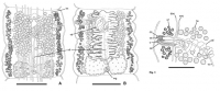
Fig. 2 Luciaella ivanovae n. g., n. sp. from Ageneiosus inermis. A, mature proglottis in dorsal view; osmoregulatory canals not drawn; B, gravid proglottis in ventral view; osmoregulatory canals and i... MoreFig. 2 Luciaella ivanovae n. g., n. sp. from Ageneiosus inermis. A, mature proglottis in dorsal view; osmoregulatory canals not drawn; B, gravid proglottis in ventral view; osmoregulatory canals and internal longitudinal musculature not drawn. Abbreviations: lm, longitudinal musculature; mg, Mehlis gland; ud, uteroduct; vd, vas deferens. Scale-bars: 500 µm. Fig. 3 Luciaella ivanovae n. g., n. sp. from Ageneiosus inermis; detail of the terminal genitalia in dorsal view. Abbreviations: c, cirrus; cc, chromophilic cells; cs, cirrus-sac; doc, dorsal osmoregulatory canal; te, testis; vc, vaginal canal; vd, vas deferens; vf, vitelline follicles; voc, ventral osmoregulatory canal; vs, vaginal sphincter. Scale-bars: 250 µm |
Photo Micrograph
|
Scanning Electron Micrograph

Fig. 4 Luciaella ivanovae n. g., n. sp. n. from Ageneiosus inermes, SEM micrographs. A, dorsoventral view of scolex indicating location of magnified surfaces shown in DJ; B, detail of the anterior lo... MoreFig. 4 Luciaella ivanovae n. g., n. sp. n. from Ageneiosus inermes, SEM micrographs. A, dorsoventral view of scolex indicating location of magnified surfaces shown in DJ; B, detail of the anterior loculus of the sucker showing the cap-peak-like structure; C, wrinkled apical surface of scolex; D, apical surface of scolex; E, luminal surface of sucker; F, marginal surface of sucker; G, non-adherent surface of sucker; H, surface of proliferation zone; I, surface of immature proglottis; J, surface of mature proglottis. Scale-bars: A, 500 µm; B, 100 µm; C, 200 µm; DJ, 2 µm |
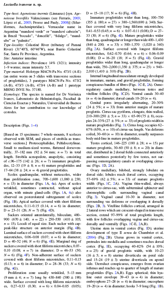
 Fig. 1 Luciaella ivanovae n. g., n. sp. from Ageneiosus inermis. A, scolex in dorsoventral view showing gland cells; B, spontaneously laid eggs; C, transverse section of mature proglottis at level of ovary; D,E, transverse sections of last immature proglottis and gravid proglottides at level of testes, showing the development of the uterus. Abbreviations: doc, dorsal osmoregulatory canal; em, embryophore; gc, gland cells; lh, larval hooks; lm, longitudinal musculature; oe, outer envelope; on, oncosphere; ov, ovary; te, testis; ut, uterus; vf, vitelline follicles; voc, ventral osmoregulatory canal. Scale-bars: A,CE, 500 µm; B, 50 µm
  Fig. 2 Luciaella ivanovae n. g., n. sp. from Ageneiosus inermis. A, mature proglottis in dorsal view; osmoregulatory canals not drawn; B, gravid proglottis in ventral view; osmoregulatory canals and internal longitudinal musculature not drawn. Abbreviations: lm, longitudinal musculature; mg, Mehlis gland; ud, uteroduct; vd, vas deferens. Scale-bars: 500 µm. Fig. 3 Luciaella ivanovae n. g., n. sp. from Ageneiosus inermis; detail of the terminal genitalia in dorsal view. Abbreviations: c, cirrus; cc, chromophilic cells; cs, cirrus-sac; doc, dorsal osmoregulatory canal; te, testis; vc, vaginal canal; vd, vas deferens; vf, vitelline follicles; voc, ventral osmoregulatory canal; vs, vaginal sphincter. Scale-bars: 250 µm
 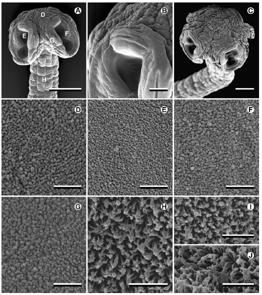 Fig. 4 Luciaella ivanovae n. g., n. sp. n. from Ageneiosus inermes, SEM micrographs. A, dorsoventral view of scolex indicating location of magnified surfaces shown in DJ; B, detail of the anterior loculus of the sucker showing the cap-peak-like structure; C, wrinkled apical surface of scolex; D, apical surface of scolex; E, luminal surface of sucker; F, marginal surface of sucker; G, non-adherent surface of sucker; H, surface of proliferation zone; I, surface of immature proglottis; J, surface of mature proglottis. Scale-bars: A, 500 µm; B, 100 µm; C, 200 µm; DJ, 2 µm

