Line Drawing 1

Figures 1-6. Pseudolacistorhynchus nanus sp. nov. from the zebra shark, Stegostoma fasciatum. 1, Entire cestode. 2,3,
Scoleces, dorso-ventral view showing variation in shape of bothria. 4, Mature se... MoreFigures 1-6. Pseudolacistorhynchus nanus sp. nov. from the zebra shark, Stegostoma fasciatum. 1, Entire cestode. 2,3,
Scoleces, dorso-ventral view showing variation in shape of bothria. 4, Mature segment. 5, Gravid segment. 6, Terminal
genital ducts, reconstructed from sections. Scale-bars: 0.1 mm. Abbreviations: c, cirrus; mg, Mehlis' gland; o, ovary; t,
testis; u, uterus; v, vagina; vd, vas deferens; vi, vitelline follicle. |
Line Drawing 2

Figures 7-13. Pseudolacistorhynchus nanus sp. nov. from the zebra shark, Stegostoma fasciatum, tentacular armature.
7. Metabasal armature, internal surface. 8. Metabasal armature, external surface; ... MoreFigures 7-13. Pseudolacistorhynchus nanus sp. nov. from the zebra shark, Stegostoma fasciatum, tentacular armature.
7. Metabasal armature, internal surface. 8. Metabasal armature, external surface; intercalary hooks shown in black. 9.
Metabasal armature, antibothrial surface. 10. Basal armature, internal surface; arrows indicate position of enlarged bill-
hooks on external surface of tentacle. 11. Basal armature, external surface. 12. Basal and metabasal armature, tentacle
slightly twisted, with internal surface of base and antibothrial surface of metabasal region; dotted line on base indicates
centre of internal surface; arrows indicate position of enlarged bill-hooks on external surface of tentacle. 13. Diagram of
arrangement of principal hooks (numerals) and intercalary hooks (letters) in metabasal region of armature; bars indicate
separation of terminal hooks from rows. Scale-bars: 0.01 mm. |
Photo Micrograph
|
Scanning Electron Micrograph
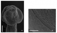
Figure 14. Pseudolacistorhynchus nanus sp. nov. from the zebra shark, Stegostoma fasciatum. Scanning electron
micrograph of scolex showing shape of bothrium. Scale-bar: 0.1 mm.
Figure 15. Pseud... MoreFigure 14. Pseudolacistorhynchus nanus sp. nov. from the zebra shark, Stegostoma fasciatum. Scanning electron
micrograph of scolex showing shape of bothrium. Scale-bar: 0.1 mm.
Figure 15. Pseudolacistorhynchus nanus sp. nov. from the zebra shark, Stegostoma fasciatum. Scanning electron
micrograph of margin of scolex showing bothrial groove and bifid microtriches. Scale-bar: 0.01 mm. |
 Figures 1-6. Pseudolacistorhynchus nanus sp. nov. from the zebra shark, Stegostoma fasciatum. 1, Entire cestode. 2,3,
Scoleces, dorso-ventral view showing variation in shape of bothria. 4, Mature segment. 5, Gravid segment. 6, Terminal
genital ducts, reconstructed from sections. Scale-bars: 0.1 mm. Abbreviations: c, cirrus; mg, Mehlis' gland; o, ovary; t,
testis; u, uterus; v, vagina; vd, vas deferens; vi, vitelline follicle.
Figures 1-6. Pseudolacistorhynchus nanus sp. nov. from the zebra shark, Stegostoma fasciatum. 1, Entire cestode. 2,3,
Scoleces, dorso-ventral view showing variation in shape of bothria. 4, Mature segment. 5, Gravid segment. 6, Terminal
genital ducts, reconstructed from sections. Scale-bars: 0.1 mm. Abbreviations: c, cirrus; mg, Mehlis' gland; o, ovary; t,
testis; u, uterus; v, vagina; vd, vas deferens; vi, vitelline follicle.  Figures 7-13. Pseudolacistorhynchus nanus sp. nov. from the zebra shark, Stegostoma fasciatum, tentacular armature.
7. Metabasal armature, internal surface. 8. Metabasal armature, external surface; intercalary hooks shown in black. 9.
Metabasal armature, antibothrial surface. 10. Basal armature, internal surface; arrows indicate position of enlarged bill-
hooks on external surface of tentacle. 11. Basal armature, external surface. 12. Basal and metabasal armature, tentacle
slightly twisted, with internal surface of base and antibothrial surface of metabasal region; dotted line on base indicates
centre of internal surface; arrows indicate position of enlarged bill-hooks on external surface of tentacle. 13. Diagram of
arrangement of principal hooks (numerals) and intercalary hooks (letters) in metabasal region of armature; bars indicate
separation of terminal hooks from rows. Scale-bars: 0.01 mm.
Figures 7-13. Pseudolacistorhynchus nanus sp. nov. from the zebra shark, Stegostoma fasciatum, tentacular armature.
7. Metabasal armature, internal surface. 8. Metabasal armature, external surface; intercalary hooks shown in black. 9.
Metabasal armature, antibothrial surface. 10. Basal armature, internal surface; arrows indicate position of enlarged bill-
hooks on external surface of tentacle. 11. Basal armature, external surface. 12. Basal and metabasal armature, tentacle
slightly twisted, with internal surface of base and antibothrial surface of metabasal region; dotted line on base indicates
centre of internal surface; arrows indicate position of enlarged bill-hooks on external surface of tentacle. 13. Diagram of
arrangement of principal hooks (numerals) and intercalary hooks (letters) in metabasal region of armature; bars indicate
separation of terminal hooks from rows. Scale-bars: 0.01 mm. 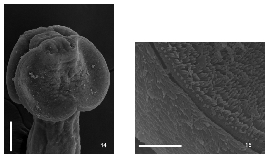 Figure 14. Pseudolacistorhynchus nanus sp. nov. from the zebra shark, Stegostoma fasciatum. Scanning electron
micrograph of scolex showing shape of bothrium. Scale-bar: 0.1 mm.
Figure 15. Pseudolacistorhynchus nanus sp. nov. from the zebra shark, Stegostoma fasciatum. Scanning electron
micrograph of margin of scolex showing bothrial groove and bifid microtriches. Scale-bar: 0.01 mm.
Figure 14. Pseudolacistorhynchus nanus sp. nov. from the zebra shark, Stegostoma fasciatum. Scanning electron
micrograph of scolex showing shape of bothrium. Scale-bar: 0.1 mm.
Figure 15. Pseudolacistorhynchus nanus sp. nov. from the zebra shark, Stegostoma fasciatum. Scanning electron
micrograph of margin of scolex showing bothrial groove and bifid microtriches. Scale-bar: 0.01 mm. 

