Line Drawing 1
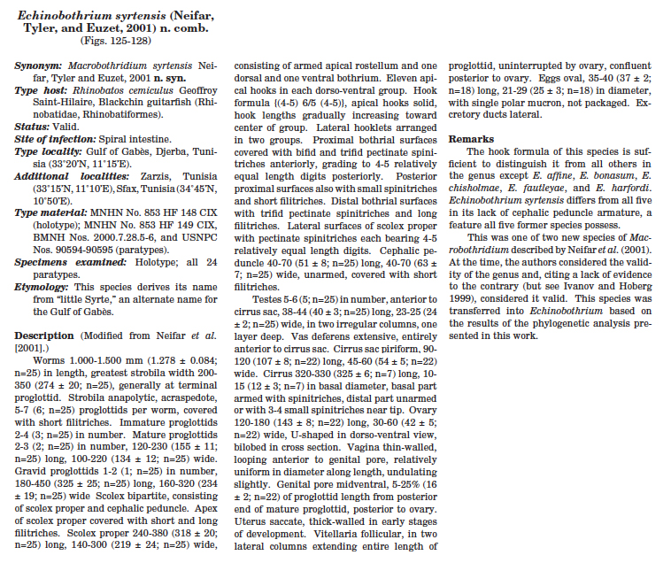
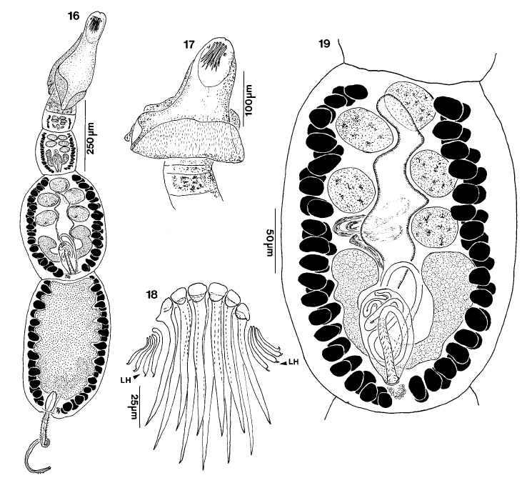
Figures 16-19. Macrobothridium syrtensis n. sp. 16. Whole worm. 17. Scolex. 18. Detail of 1 dorsoventral group of rostellar armature. 19. mature segment. LH, lateral hooklet. |
Line Drawing 2
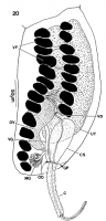
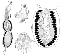
Figure 20. Gravid segment of Macrobothridium syrtensis n. sp., lateral view. C, cirrus; CS ,cirrus sac; GP genital pore; MG, Mehlis gland; OD, ovidcut; OV, ovary; UT, uterus; VD, vas deferens; VF, vit... MoreFigure 20. Gravid segment of Macrobothridium syrtensis n. sp., lateral view. C, cirrus; CS ,cirrus sac; GP genital pore; MG, Mehlis gland; OD, ovidcut; OV, ovary; UT, uterus; VD, vas deferens; VF, vitellien follicle; VG, vagina. |
Photo Micrograph
|
Scanning Electron Micrograph
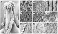
Figures 21-30. Scanning electron micrographs of Macrobothridium syrtensis n. sp. 21. Scolex. Arrow indicates border between scolex proper and cephalic peduncle. 22. Short and long filitriches on apex... MoreFigures 21-30. Scanning electron micrographs of Macrobothridium syrtensis n. sp. 21. Scolex. Arrow indicates border between scolex proper and cephalic peduncle. 22. Short and long filitriches on apex of scolex proper. 23. Pectinate spinitriches on anterior proximal bothrial surfaces. 24. Pectinate spinitriches, small spinitriches, and short filitriches on posterior proximal bothrial surfaces. 25 .Long filitriches and pectinate spinitriches on dorsal surace of scolex, anterior to hooks. 26. Long filitriches and pectinate spinitriches on medial distal bothrial surfaces. 27. Long filitriches on lateral distal bothrial surfaces. 28. Border between proximal and distal bothrial surfaces. 29. Pectinate spinitriches on lateral portion of scolex proper, between bothria. 30. Short filitriches on strobila. Scale bar in Figure 21, 50µm. Scale bar in Figure 28, 2 µm. Scale bars in Figures 22-27 and 29-30, 1 µm. |
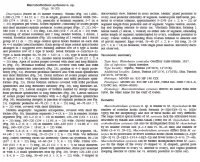
 Figures 16-19. Macrobothridium syrtensis n. sp. 16. Whole worm. 17. Scolex. 18. Detail of 1 dorsoventral group of rostellar armature. 19. mature segment. LH, lateral hooklet.
  Figure 20. Gravid segment of Macrobothridium syrtensis n. sp., lateral view. C, cirrus; CS ,cirrus sac; GP genital pore; MG, Mehlis gland; OD, ovidcut; OV, ovary; UT, uterus; VD, vas deferens; VF, vitellien follicle; VG, vagina.
  Figures 21-30. Scanning electron micrographs of Macrobothridium syrtensis n. sp. 21. Scolex. Arrow indicates border between scolex proper and cephalic peduncle. 22. Short and long filitriches on apex of scolex proper. 23. Pectinate spinitriches on anterior proximal bothrial surfaces. 24. Pectinate spinitriches, small spinitriches, and short filitriches on posterior proximal bothrial surfaces. 25 .Long filitriches and pectinate spinitriches on dorsal surace of scolex, anterior to hooks. 26. Long filitriches and pectinate spinitriches on medial distal bothrial surfaces. 27. Long filitriches on lateral distal bothrial surfaces. 28. Border between proximal and distal bothrial surfaces. 29. Pectinate spinitriches on lateral portion of scolex proper, between bothria. 30. Short filitriches on strobila. Scale bar in Figure 21, 50µm. Scale bar in Figure 28, 2 µm. Scale bars in Figures 22-27 and 29-30, 1 µm.