Line Drawing 1
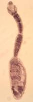
FIGURES 2130. Echinobothrium rayallemangi n. sp. 21. Whole worm. 22. Scolex. 23. Detail of 1 dorsoventral group of apical hooks. 24.
Detail of 1 group of lateral hooklets. 25. Detail of cephalic pe... MoreFIGURES 2130. Echinobothrium rayallemangi n. sp. 21. Whole worm. 22. Scolex. 23. Detail of 1 dorsoventral group of apical hooks. 24.
Detail of 1 group of lateral hooklets. 25. Detail of cephalic peduncle spine. 26. Mature segment. 27. Cross section through segment at level indicated by arrow at 27 in Figure 26. 28. Cross section through segment at level indicated by arrow at 28 in Figure 26. 29. Lateral view of
terminal genitalia. 30. Eggs. AH, apical hook; C, cirrus; CP, cephalic peduncle; GP, genital pore; LH, lateral hooklets; MG, Mehlis gland; OD, oviduct; OV, ovar y; SP, scolex proper; T, testis; UD, uterine duct; UT, uterus; VD, vas deferens; VF, vitelline follicle; VG, vagina. |
Line Drawing 2
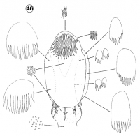
FIGURE 46. Summar y of distributions of various microtrich forms on scolex of Echinobothrium rayallemangi n. sp. All microtriches are drawn to same scale. |
Photo Micrograph
|
Scanning Electron Micrograph
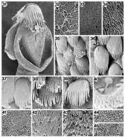
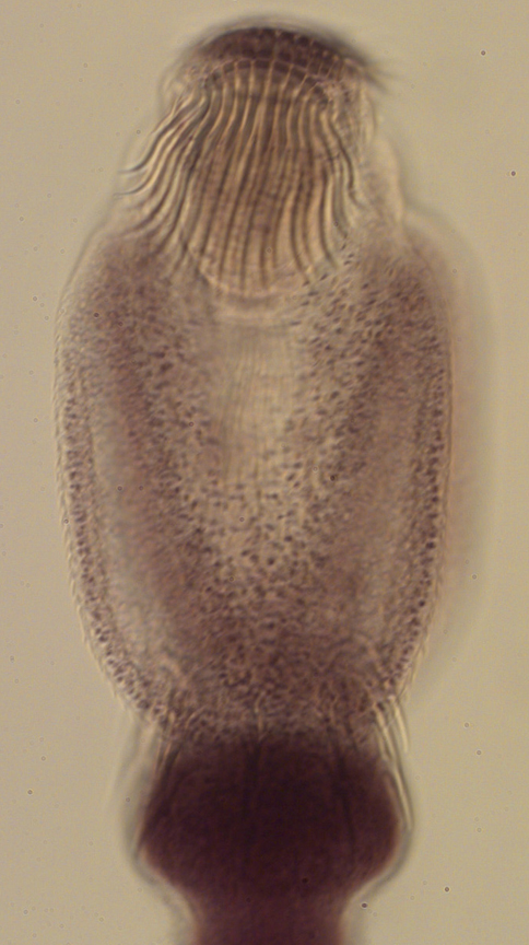
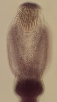
FIGURES 31 45. Scanning electron micrographs of Echinobothrium rayallemangi n. sp. 31. Scolex proper. 32. Apex of scolex. 33. Dorsal region of scolex anterior to apical hooks. 34. Lateral portion of ... MoreFIGURES 31 45. Scanning electron micrographs of Echinobothrium rayallemangi n. sp. 31. Scolex proper. 32. Apex of scolex. 33. Dorsal region of scolex anterior to apical hooks. 34. Lateral portion of scolex, anterior to lateral hooklets. 35. Anterior proximal bothrial surface. 36. Proximal bothrial surface, halfway along length, showing cilium (arrow). 37. Posterior proximal bothrial surface. 38. Anterior distal bothrial
surface. 39. Distal bothrial surface, halfway along length. 40. Posterior edge of bothrium, showing microtriches on both proximal and distal surface. 41. Medial distal bothrial surface. 42. Anterior lateral portion of scolex proper. 43. Posterior lateral portion of scolex proper. 44. Neck. 45. Strobila. Scale bar in Figure 31, 40 um. Scale bars in Figures 3239, 41 45, 1 um. Scale bar in Figure 40, 5 um. |
 FIGURES 2130. Echinobothrium rayallemangi n. sp. 21. Whole worm. 22. Scolex. 23. Detail of 1 dorsoventral group of apical hooks. 24.
Detail of 1 group of lateral hooklets. 25. Detail of cephalic peduncle spine. 26. Mature segment. 27. Cross section through segment at level indicated by arrow at 27 in Figure 26. 28. Cross section through segment at level indicated by arrow at 28 in Figure 26. 29. Lateral view of
terminal genitalia. 30. Eggs. AH, apical hook; C, cirrus; CP, cephalic peduncle; GP, genital pore; LH, lateral hooklets; MG, Mehlis gland; OD, oviduct; OV, ovar y; SP, scolex proper; T, testis; UD, uterine duct; UT, uterus; VD, vas deferens; VF, vitelline follicle; VG, vagina.
FIGURES 2130. Echinobothrium rayallemangi n. sp. 21. Whole worm. 22. Scolex. 23. Detail of 1 dorsoventral group of apical hooks. 24.
Detail of 1 group of lateral hooklets. 25. Detail of cephalic peduncle spine. 26. Mature segment. 27. Cross section through segment at level indicated by arrow at 27 in Figure 26. 28. Cross section through segment at level indicated by arrow at 28 in Figure 26. 29. Lateral view of
terminal genitalia. 30. Eggs. AH, apical hook; C, cirrus; CP, cephalic peduncle; GP, genital pore; LH, lateral hooklets; MG, Mehlis gland; OD, oviduct; OV, ovar y; SP, scolex proper; T, testis; UD, uterine duct; UT, uterus; VD, vas deferens; VF, vitelline follicle; VG, vagina. 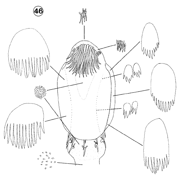 FIGURE 46. Summar y of distributions of various microtrich forms on scolex of Echinobothrium rayallemangi n. sp. All microtriches are drawn to same scale.
  FIGURES 31 45. Scanning electron micrographs of Echinobothrium rayallemangi n. sp. 31. Scolex proper. 32. Apex of scolex. 33. Dorsal region of scolex anterior to apical hooks. 34. Lateral portion of scolex, anterior to lateral hooklets. 35. Anterior proximal bothrial surface. 36. Proximal bothrial surface, halfway along length, showing cilium (arrow). 37. Posterior proximal bothrial surface. 38. Anterior distal bothrial
surface. 39. Distal bothrial surface, halfway along length. 40. Posterior edge of bothrium, showing microtriches on both proximal and distal surface. 41. Medial distal bothrial surface. 42. Anterior lateral portion of scolex proper. 43. Posterior lateral portion of scolex proper. 44. Neck. 45. Strobila. Scale bar in Figure 31, 40 um. Scale bars in Figures 3239, 41 45, 1 um. Scale bar in Figure 40, 5 um.
FIGURES 31 45. Scanning electron micrographs of Echinobothrium rayallemangi n. sp. 31. Scolex proper. 32. Apex of scolex. 33. Dorsal region of scolex anterior to apical hooks. 34. Lateral portion of scolex, anterior to lateral hooklets. 35. Anterior proximal bothrial surface. 36. Proximal bothrial surface, halfway along length, showing cilium (arrow). 37. Posterior proximal bothrial surface. 38. Anterior distal bothrial
surface. 39. Distal bothrial surface, halfway along length. 40. Posterior edge of bothrium, showing microtriches on both proximal and distal surface. 41. Medial distal bothrial surface. 42. Anterior lateral portion of scolex proper. 43. Posterior lateral portion of scolex proper. 44. Neck. 45. Strobila. Scale bar in Figure 31, 40 um. Scale bars in Figures 3239, 41 45, 1 um. Scale bar in Figure 40, 5 um.