Cestode Scientific Name
| Species ID | 5735 |
|---|---|
| Order | Diphyllidea |
| Family | |
| Subfamily | |
| Genus | Echinobothrium |
| Species | clavatum |
| Authority | Probert & Stobart, 1989 |
| Taxonomic Status | Valid |
| Valid Name | |
| Synonyms | |
| Genus Record | No |
| Type Species | No |
| Verified | Yes |
| Verified By | R. Kuchta, V. A. Ivanov |
| Citation(s) |
Probert, A. J. and B. Stobart. 1989. Echinobothrium clavatum n. sp. (Cestoda, Diphyllidea) from the spiral valve of Raja clavata L., 1758, including a note on its ultrastructure and a key to species of the genus.. Systematic Parasitology 13: 71-79. (349) Download PDF |
| Redescription | |
| Scientific Name Notes |
Record Data
| Date (MM/DD/YYYY) | Action | User Name |
|---|---|---|
| 09/30/2009 | Created | V. Lopez , V.A, V.A. Ivanov, R. |
| 08/20/2014 | Modified | |
| 02/24/2016 | Modified | B. Barbeau |
| 06/17/2016 | Modified | B. Barbeau |
| 08/23/2016 | Modified | R. Kuchta |
Type Host
| Host Class | Chondrichthyes | ||||||
|---|---|---|---|---|---|---|---|
| Host Order | Rajiformes | ||||||
| Host Family | Rajidae | ||||||
|
Type Host (Literal) |
|
||||||
|
Type Host (Valid) |
|
||||||
| Additional Host(s) | |||||||
| Site in Host | spiral valve | ||||||
| Host Notes |
Type Locality
| Country | North Wales |
|---|---|
| Body of Water | Irish Sea |
| Island(s) | |
| City/Region | off Anglesey |
| Coordinates | |
| DD Latitude | |
| DD Longitude | |
| Additional Localities | |
| Locality Notes |
Specimens
| Type Material | BMNH No. 1988.6.1.1-3 (syntypes) |
|---|---|
| Total Number of Type Specimens | Based on the measurement of 11 specimens |
| Voucher Material | |
| Specimen Notes | Tyler (2006) designated specimen BMNH No. 1988.6.1.1 as a lectotype |
Data are given as in original description unless otherwise indicated.

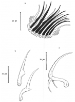
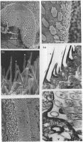
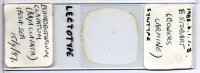
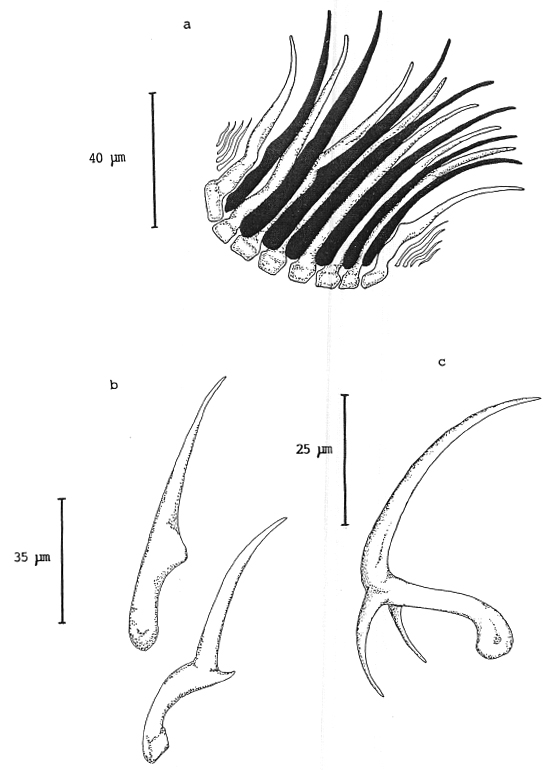 Fig. 1. Echinobothrium clavatum n. sp. a. The apical hooks. b. The shape of the upper and lower apical hooks. c. The shape of the
spines of the cephalic peduncle.
Fig. 1. Echinobothrium clavatum n. sp. a. The apical hooks. b. The shape of the upper and lower apical hooks. c. The shape of the
spines of the cephalic peduncle.  Fig. 2. Echinobothriurn clavaturn n. sp. a. Scanning electron micrograph of the undersurface of the bothridium (b) showing the
scales (sc) and the outer surface covered with spines (s). b. S.E.M. of the edge of the bothridium showing the four cusped scales
(sc) on the undersurface and the spines (s) on the outer edge. c. S.E.M. showing the dense mat of spines on the outer surface of the
bothridium. d. T.E.M. showing the shape of the bothridial spines. Note the T-shaped base of the bothridial spine inserted in the
tegument. x 15,000. e. T.E.M. of the junction between the parasite and the host microvilli (hm). Note the highly vacuolate
tegument (v) and the intimate contact (0) between the parasite and the host microvilli the tips of which are electron dense. x6,000.
f. T.E.M. of the host parasite interface showing the host microvilli (m), the vacuolated tegument of the parasite (v), and
pinocytotic vesicle (p arrow). x 60,000.
Fig. 2. Echinobothriurn clavaturn n. sp. a. Scanning electron micrograph of the undersurface of the bothridium (b) showing the
scales (sc) and the outer surface covered with spines (s). b. S.E.M. of the edge of the bothridium showing the four cusped scales
(sc) on the undersurface and the spines (s) on the outer edge. c. S.E.M. showing the dense mat of spines on the outer surface of the
bothridium. d. T.E.M. showing the shape of the bothridial spines. Note the T-shaped base of the bothridial spine inserted in the
tegument. x 15,000. e. T.E.M. of the junction between the parasite and the host microvilli (hm). Note the highly vacuolate
tegument (v) and the intimate contact (0) between the parasite and the host microvilli the tips of which are electron dense. x6,000.
f. T.E.M. of the host parasite interface showing the host microvilli (m), the vacuolated tegument of the parasite (v), and
pinocytotic vesicle (p arrow). x 60,000. 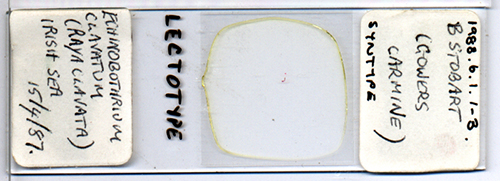 BMNH No. 1988.6.1-3, lectotype slide
 Best viewed in Firefox


