Line Drawing 1
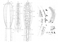
Text-figs. 1-3. Ditrachybothridium macrocephalum gen.nov., sp.nov.
1. Scolex, dorsal or ventral view, showing bothridium with marginal spines and the nervous system. 2. Scolex, lateral view, showing ... MoreText-figs. 1-3. Ditrachybothridium macrocephalum gen.nov., sp.nov.
1. Scolex, dorsal or ventral view, showing bothridium with marginal spines and the nervous system. 2. Scolex, lateral view, showing nervous and excretory systems. 3. Scolex, dorsal view, showing dorsal half of excretory system. Text-figs. 4-9. Ditrachybothridium macrocephalum gen.nov., sp.nov. 4. Scolex, lateral view, showing outer convex face of the dorsal and ventral bothridia rolled over, exposing the rows of backwardly directed spines. 5(a), (b) and (c). Arrangement of spines on the anterior, middle and posterior regions of the outer convex face of the bothridium, surface view. 6(a), (b) and (c). Arrangement of spines on the anterior, middle and posterior regions of the margin of the bothridium, surface view. 7. Series of ten spines taken at intervals from the anterior to the posterior ends of the margin of the bothridium, showing progressive decrease in size. 8. Series of five spines selected from those of one row on the outer convex surface of the bothridium, showing progressive decrease in size from the margin inwards. 9. Transverse section through the free margin of one bothridium, showing arrangement of spines on the outer convex surface. Keys to Lettering in Figures (All drawings are semi-diagrammatic) a.l. anterior loop of excretory vessel, a.n. anterior nerve, a.o. apical organ, b. bothridium, b.n. bothridial nerve, c.c. cuticle-secreting cells, c.g. cerebral ganglion, c.s. cirrus sac, cu. cuticle, d.e.v. dorsal excretory vessel, d.v.m. dorso-ventral muscles, e.n.s. excretory network in scolex, e.p. excretory pore, g.p. genital pore, l.m. longitudinal muscles, l.n. lateral nerve, m.d. muscular depression around excretory pore, m.g. Mehlis's gland, n. neck, ov. ovary, ovd. oviduct, p. parenchyma, p.n. posterior nerve, r.m. radial muscles, s. scolex, sp. spine, t. testis, t.m. transverse muscles, ut. uterus, v. velum, v.e.v. ventral excretory vessel, vag. vagina, v.d. vas deferens, vit. yolk follicle, vit.d. yolk duct. |
Line Drawing 2
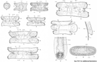
Text-figs. 10-16 Ditrachybothridium macrocephalum gen.nov., sp.nov. 10. Transverse section through tip of scolex, showing feebly developed apical organ. 11. Transverse section through scolex, showing ... MoreText-figs. 10-16 Ditrachybothridium macrocephalum gen.nov., sp.nov. 10. Transverse section through tip of scolex, showing feebly developed apical organ. 11. Transverse section through scolex, showing beginnings of bothridia and dorso-ventral excretory loop on either side of the apical organ. 12. Transverse section through scolex immediately behind apical organ and front of brain showing bothridia with spines, excretory vessels and the transverse, dorso-vetnral, radial and longitudinal muscles. 13. Transverse section through the scolex, showing cerebral ganglia united by transverse commissure and the musculature. 14. Transverse section through the scolex immediately behind the cerebral ganglia, showing the lateral nerves and the better developed musculature in the region. 15. Transverse section through the scolex, showing the bothridial nerves arising from the lateral nerves; the bothridia, musculature and excretory vessels as before. 16. Transverse section through the scolex, showing, mainly, extension of the radial muscles towards the centre. Text-figs. 17-21. Ditrachybothridium macrocephalum gen.nov., sp.nov. 17. Transverse section through the scolex, showing decrease in prominence of the bothridia, change in direction of the bothridial nerves as they arise, the branching of the excretory vessels and the poorly developed musculature. 18. Transverse section through the posterior end of the scolex, showing further narrowing of the bothridia and consequently more prominent lateral region of the scolex. 19. Portion of sagittal section through margin of bothridium, showing bundle of longitudinal muscles. 20 Transverse section through neck, showing very weakly developed muscles, excretory vessels, lateral nerves and the cells filling a large part of the parenchyma. 21. Excretory pores in terminal segment, ventral view. Keys to Lettering in Figures (All drawings are semi-diagrammatic) a.l. anterior loop of excretory vessel, a.n. anterior nerve, a.o. apical organ, b. bothridium, b.n. bothridial nerve, c.c. cuticle-secreting cells, c.g. cerebral ganglion, c.s. cirrus sac, cu. cuticle, d.e.v. dorsal excretory vessel, d.v.m. dorso-ventral muscles, e.n.s. excretory network in scolex, e.p. excretory pore, g.p. genital pore, l.m. longitudinal muscles, l.n. lateral nerve, m.d. muscular depression around excretory pore, m.g. Mehlis's gland, n. neck, ov. ovary, ovd. oviduct, p. parenchyma, p.n. posterior nerve, r.m. radial muscles, s. scolex, sp. spine, t. testis, t.m. transverse muscles, ut. uterus, v. velum, v.e.v. ventral excretory vessel, vag. vagina, v.d. vas deferens, vit. yolk follicle, vit.d. yolk duct. [Text-figs 22-27 not shown] |
Photo Micrograph
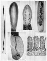
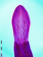
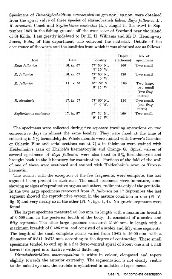
Fig. 1 Entire worm. Fig. 2. Scolex, dorsal or ventral view, showing spines along margin of bothridium. Fig. 3. Contracted scolex, showing some of the excretory vessels. Fig. 4. Last segment of complet... MoreFig. 1 Entire worm. Fig. 2. Scolex, dorsal or ventral view, showing spines along margin of bothridium. Fig. 3. Contracted scolex, showing some of the excretory vessels. Fig. 4. Last segment of complete worm (fig. 1), showing testes and immature female genitalia. Fig. 5. Last segment of another complete worm, showing mature genitalia (see Text-fig. 23). Fig. 6. Vertical section through wall of spiral valve of Raja fullonica to show villi. |
Scanning Electron Micrograph
|


 Text-figs. 1-3. Ditrachybothridium macrocephalum gen.nov., sp.nov.
1. Scolex, dorsal or ventral view, showing bothridium with marginal spines and the nervous system. 2. Scolex, lateral view, showing nervous and excretory systems. 3. Scolex, dorsal view, showing dorsal half of excretory system. Text-figs. 4-9. Ditrachybothridium macrocephalum gen.nov., sp.nov. 4. Scolex, lateral view, showing outer convex face of the dorsal and ventral bothridia rolled over, exposing the rows of backwardly directed spines. 5(a), (b) and (c). Arrangement of spines on the anterior, middle and posterior regions of the outer convex face of the bothridium, surface view. 6(a), (b) and (c). Arrangement of spines on the anterior, middle and posterior regions of the margin of the bothridium, surface view. 7. Series of ten spines taken at intervals from the anterior to the posterior ends of the margin of the bothridium, showing progressive decrease in size. 8. Series of five spines selected from those of one row on the outer convex surface of the bothridium, showing progressive decrease in size from the margin inwards. 9. Transverse section through the free margin of one bothridium, showing arrangement of spines on the outer convex surface. Keys to Lettering in Figures (All drawings are semi-diagrammatic) a.l. anterior loop of excretory vessel, a.n. anterior nerve, a.o. apical organ, b. bothridium, b.n. bothridial nerve, c.c. cuticle-secreting cells, c.g. cerebral ganglion, c.s. cirrus sac, cu. cuticle, d.e.v. dorsal excretory vessel, d.v.m. dorso-ventral muscles, e.n.s. excretory network in scolex, e.p. excretory pore, g.p. genital pore, l.m. longitudinal muscles, l.n. lateral nerve, m.d. muscular depression around excretory pore, m.g. Mehlis's gland, n. neck, ov. ovary, ovd. oviduct, p. parenchyma, p.n. posterior nerve, r.m. radial muscles, s. scolex, sp. spine, t. testis, t.m. transverse muscles, ut. uterus, v. velum, v.e.v. ventral excretory vessel, vag. vagina, v.d. vas deferens, vit. yolk follicle, vit.d. yolk duct.
Text-figs. 1-3. Ditrachybothridium macrocephalum gen.nov., sp.nov.
1. Scolex, dorsal or ventral view, showing bothridium with marginal spines and the nervous system. 2. Scolex, lateral view, showing nervous and excretory systems. 3. Scolex, dorsal view, showing dorsal half of excretory system. Text-figs. 4-9. Ditrachybothridium macrocephalum gen.nov., sp.nov. 4. Scolex, lateral view, showing outer convex face of the dorsal and ventral bothridia rolled over, exposing the rows of backwardly directed spines. 5(a), (b) and (c). Arrangement of spines on the anterior, middle and posterior regions of the outer convex face of the bothridium, surface view. 6(a), (b) and (c). Arrangement of spines on the anterior, middle and posterior regions of the margin of the bothridium, surface view. 7. Series of ten spines taken at intervals from the anterior to the posterior ends of the margin of the bothridium, showing progressive decrease in size. 8. Series of five spines selected from those of one row on the outer convex surface of the bothridium, showing progressive decrease in size from the margin inwards. 9. Transverse section through the free margin of one bothridium, showing arrangement of spines on the outer convex surface. Keys to Lettering in Figures (All drawings are semi-diagrammatic) a.l. anterior loop of excretory vessel, a.n. anterior nerve, a.o. apical organ, b. bothridium, b.n. bothridial nerve, c.c. cuticle-secreting cells, c.g. cerebral ganglion, c.s. cirrus sac, cu. cuticle, d.e.v. dorsal excretory vessel, d.v.m. dorso-ventral muscles, e.n.s. excretory network in scolex, e.p. excretory pore, g.p. genital pore, l.m. longitudinal muscles, l.n. lateral nerve, m.d. muscular depression around excretory pore, m.g. Mehlis's gland, n. neck, ov. ovary, ovd. oviduct, p. parenchyma, p.n. posterior nerve, r.m. radial muscles, s. scolex, sp. spine, t. testis, t.m. transverse muscles, ut. uterus, v. velum, v.e.v. ventral excretory vessel, vag. vagina, v.d. vas deferens, vit. yolk follicle, vit.d. yolk duct.  Text-figs. 10-16 Ditrachybothridium macrocephalum gen.nov., sp.nov. 10. Transverse section through tip of scolex, showing feebly developed apical organ. 11. Transverse section through scolex, showing beginnings of bothridia and dorso-ventral excretory loop on either side of the apical organ. 12. Transverse section through scolex immediately behind apical organ and front of brain showing bothridia with spines, excretory vessels and the transverse, dorso-vetnral, radial and longitudinal muscles. 13. Transverse section through the scolex, showing cerebral ganglia united by transverse commissure and the musculature. 14. Transverse section through the scolex immediately behind the cerebral ganglia, showing the lateral nerves and the better developed musculature in the region. 15. Transverse section through the scolex, showing the bothridial nerves arising from the lateral nerves; the bothridia, musculature and excretory vessels as before. 16. Transverse section through the scolex, showing, mainly, extension of the radial muscles towards the centre. Text-figs. 17-21. Ditrachybothridium macrocephalum gen.nov., sp.nov. 17. Transverse section through the scolex, showing decrease in prominence of the bothridia, change in direction of the bothridial nerves as they arise, the branching of the excretory vessels and the poorly developed musculature. 18. Transverse section through the posterior end of the scolex, showing further narrowing of the bothridia and consequently more prominent lateral region of the scolex. 19. Portion of sagittal section through margin of bothridium, showing bundle of longitudinal muscles. 20 Transverse section through neck, showing very weakly developed muscles, excretory vessels, lateral nerves and the cells filling a large part of the parenchyma. 21. Excretory pores in terminal segment, ventral view. Keys to Lettering in Figures (All drawings are semi-diagrammatic) a.l. anterior loop of excretory vessel, a.n. anterior nerve, a.o. apical organ, b. bothridium, b.n. bothridial nerve, c.c. cuticle-secreting cells, c.g. cerebral ganglion, c.s. cirrus sac, cu. cuticle, d.e.v. dorsal excretory vessel, d.v.m. dorso-ventral muscles, e.n.s. excretory network in scolex, e.p. excretory pore, g.p. genital pore, l.m. longitudinal muscles, l.n. lateral nerve, m.d. muscular depression around excretory pore, m.g. Mehlis's gland, n. neck, ov. ovary, ovd. oviduct, p. parenchyma, p.n. posterior nerve, r.m. radial muscles, s. scolex, sp. spine, t. testis, t.m. transverse muscles, ut. uterus, v. velum, v.e.v. ventral excretory vessel, vag. vagina, v.d. vas deferens, vit. yolk follicle, vit.d. yolk duct. [Text-figs 22-27 not shown]
  Fig. 1 Entire worm. Fig. 2. Scolex, dorsal or ventral view, showing spines along margin of bothridium. Fig. 3. Contracted scolex, showing some of the excretory vessels. Fig. 4. Last segment of complete worm (fig. 1), showing testes and immature female genitalia. Fig. 5. Last segment of another complete worm, showing mature genitalia (see Text-fig. 23). Fig. 6. Vertical section through wall of spiral valve of Raja fullonica to show villi.
 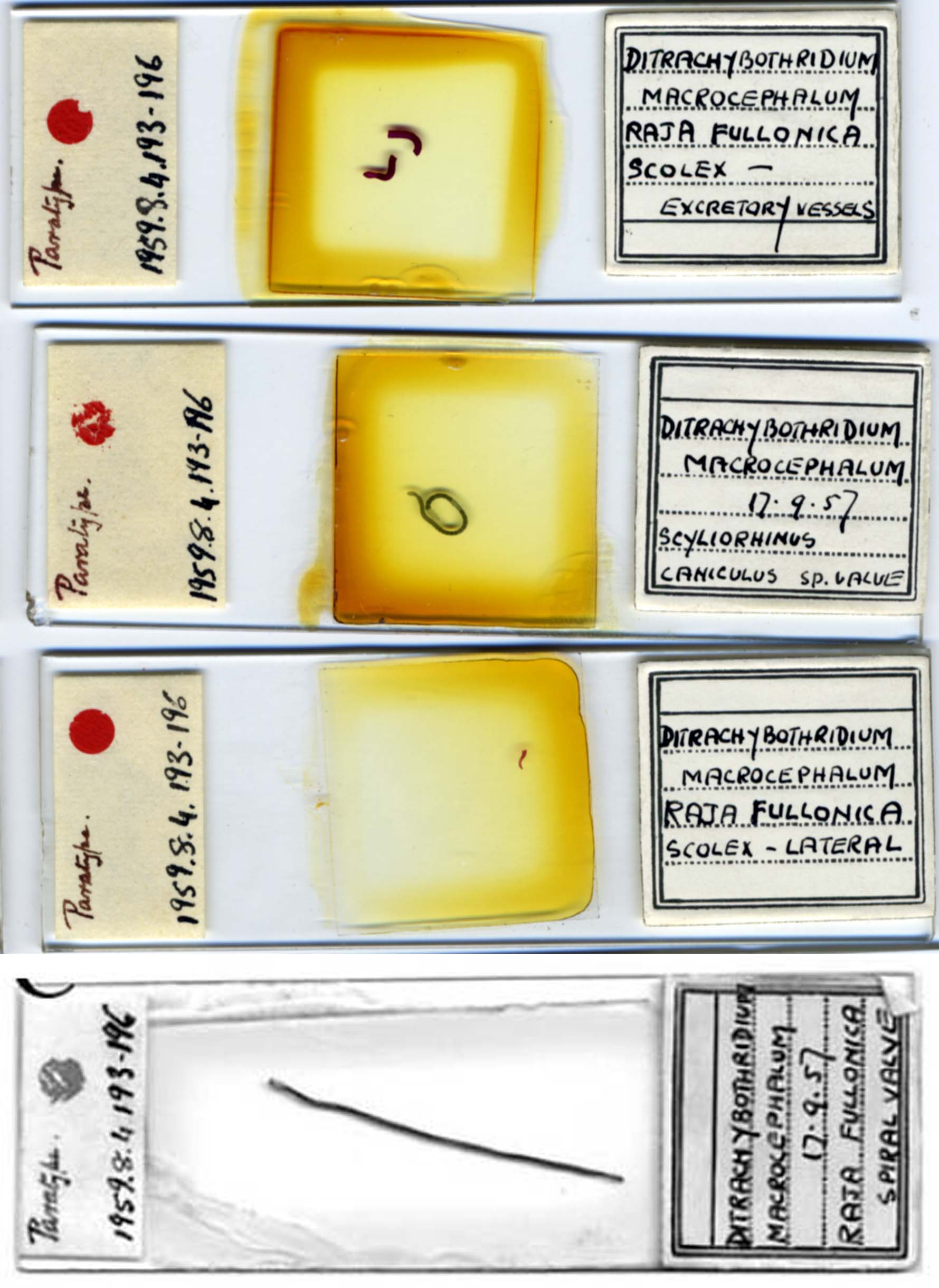 BMNH No. 1959.8.4.193-196 (paratype)
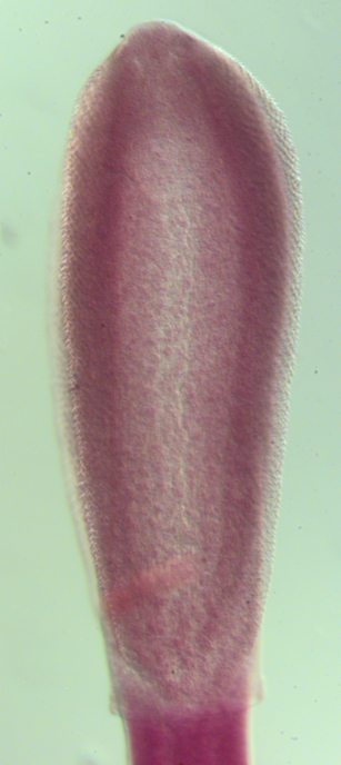
 BMNH No. 1959.8.4.193-196 (paratype)
  BMNH No. 1959.8.4.193-196 (paratype)

