Line Drawing 1
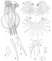
FIGURES 16. E. diamanti n. sp. (1) Scolex proper, bar= 100 µm. (2) Apical hooks, lateral hooklets, and a subset of spines from the corona, lateral view, bar= 20 µm. (3) Apical hooks (1 dorsoventral ... MoreFIGURES 16. E. diamanti n. sp. (1) Scolex proper, bar= 100 µm. (2) Apical hooks, lateral hooklets, and a subset of spines from the corona, lateral view, bar= 20 µm. (3) Apical hooks (1 dorsoventral group) and lateral hooklets, apical view, bar = 20 µm (114 anterior row, 1'13' posterior row). (4) Detail of small spines from corona posterior to apical hooks, bar = 10 µm. (5) Detail of apical hooks, a Hook from anterior row. b Hook from posterior row, bar= 10 µm. (6) Detail of spines on cephalic peduncle, a Anteriormost spines. b Spine from middle zone. c Posteriormost spines, bar = 20 µm. |
Line Drawing 2
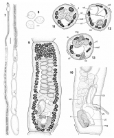
FIGURES 713. E. diamanti n. sp. (7) Entire worm, bar= 500 µm. (8) Detail of eggs, bar= 20 µm. (9) Mature proglottid, ventral view (vitelline
follicles are only partially drawn to allow the view of i... MoreFIGURES 713. E. diamanti n. sp. (7) Entire worm, bar= 500 µm. (8) Detail of eggs, bar= 20 µm. (9) Mature proglottid, ventral view (vitelline
follicles are only partially drawn to allow the view of internal organs), bar = 100 µm. (10) Detail of genitalia, lateral view (vitelline follicles are not drawn), bar = 150 µm. (11) Cross section of mature proglottid at level of testes, bar = 50 µm. (12) Cross section of mature proglottid at level of cirrus sac, bar = 50 µm. (13) Cross section of mature proglottid at level of ovarian isthmus, bar = 50 µm. Abbreviations: cs, cirrus sac; mg, Mehlis gland; o, ovary; t, testis; ud, uterine duct; ut, uterus; vd, vas deferens; vf, vitelline follicle; vod, ventral osmoregulatory duct; vs, vaginal sphincter |
Photo Micrograph
|
Scanning Electron Micrograph
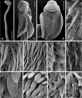
FIGURES 1425. E. diamanti n. sp., scanning electron micrographs. (14) Complete scolex consisting of scolex proper and cephalic peduncle, bar= 500 µm. (15) Scolex proper, lateral view, bar= 100 µm. (1... MoreFIGURES 1425. E. diamanti n. sp., scanning electron micrographs. (14) Complete scolex consisting of scolex proper and cephalic peduncle, bar= 500 µm. (15) Scolex proper, lateral view, bar= 100 µm. (16) Scolex proper, dorsal/ventral view, bar =100 µm. (17) Spines from corona at its posterior margin, note the surface of the tegument is devoid of microtriches, bar= 4 µm. (18) Detail of microtriches on distal bothrial surface
(anterior region), bar = 2 µm. (19) Distal bothrial surface (anterior region), bar = 2.5 µm. (20) Lateral margin of bothria showing border between distal and proximal surfaces (anterior region), bar = 2 µm. (21) Surface of cirrus, bar = 2 µm. (22) Detail of microtriches on proximal bothrial surface, bar = 2 µm. (23) Proximal bothrial surface (anterior region), bar = 2 µm. (24) Surface of proliferation zone, bar = 1 µm. (25) Surface of mature proglottids, bar= 1 µm. |

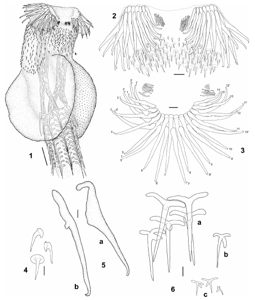 FIGURES 16. E. diamanti n. sp. (1) Scolex proper, bar= 100 µm. (2) Apical hooks, lateral hooklets, and a subset of spines from the corona, lateral view, bar= 20 µm. (3) Apical hooks (1 dorsoventral group) and lateral hooklets, apical view, bar = 20 µm (114 anterior row, 1'13' posterior row). (4) Detail of small spines from corona posterior to apical hooks, bar = 10 µm. (5) Detail of apical hooks, a Hook from anterior row. b Hook from posterior row, bar= 10 µm. (6) Detail of spines on cephalic peduncle, a Anteriormost spines. b Spine from middle zone. c Posteriormost spines, bar = 20 µm.
  FIGURES 713. E. diamanti n. sp. (7) Entire worm, bar= 500 µm. (8) Detail of eggs, bar= 20 µm. (9) Mature proglottid, ventral view (vitelline
follicles are only partially drawn to allow the view of internal organs), bar = 100 µm. (10) Detail of genitalia, lateral view (vitelline follicles are not drawn), bar = 150 µm. (11) Cross section of mature proglottid at level of testes, bar = 50 µm. (12) Cross section of mature proglottid at level of cirrus sac, bar = 50 µm. (13) Cross section of mature proglottid at level of ovarian isthmus, bar = 50 µm. Abbreviations: cs, cirrus sac; mg, Mehlis gland; o, ovary; t, testis; ud, uterine duct; ut, uterus; vd, vas deferens; vf, vitelline follicle; vod, ventral osmoregulatory duct; vs, vaginal sphincter
FIGURES 713. E. diamanti n. sp. (7) Entire worm, bar= 500 µm. (8) Detail of eggs, bar= 20 µm. (9) Mature proglottid, ventral view (vitelline
follicles are only partially drawn to allow the view of internal organs), bar = 100 µm. (10) Detail of genitalia, lateral view (vitelline follicles are not drawn), bar = 150 µm. (11) Cross section of mature proglottid at level of testes, bar = 50 µm. (12) Cross section of mature proglottid at level of cirrus sac, bar = 50 µm. (13) Cross section of mature proglottid at level of ovarian isthmus, bar = 50 µm. Abbreviations: cs, cirrus sac; mg, Mehlis gland; o, ovary; t, testis; ud, uterine duct; ut, uterus; vd, vas deferens; vf, vitelline follicle; vod, ventral osmoregulatory duct; vs, vaginal sphincter  FIGURES 1425. E. diamanti n. sp., scanning electron micrographs. (14) Complete scolex consisting of scolex proper and cephalic peduncle, bar= 500 µm. (15) Scolex proper, lateral view, bar= 100 µm. (16) Scolex proper, dorsal/ventral view, bar =100 µm. (17) Spines from corona at its posterior margin, note the surface of the tegument is devoid of microtriches, bar= 4 µm. (18) Detail of microtriches on distal bothrial surface
(anterior region), bar = 2 µm. (19) Distal bothrial surface (anterior region), bar = 2.5 µm. (20) Lateral margin of bothria showing border between distal and proximal surfaces (anterior region), bar = 2 µm. (21) Surface of cirrus, bar = 2 µm. (22) Detail of microtriches on proximal bothrial surface, bar = 2 µm. (23) Proximal bothrial surface (anterior region), bar = 2 µm. (24) Surface of proliferation zone, bar = 1 µm. (25) Surface of mature proglottids, bar= 1 µm.
FIGURES 1425. E. diamanti n. sp., scanning electron micrographs. (14) Complete scolex consisting of scolex proper and cephalic peduncle, bar= 500 µm. (15) Scolex proper, lateral view, bar= 100 µm. (16) Scolex proper, dorsal/ventral view, bar =100 µm. (17) Spines from corona at its posterior margin, note the surface of the tegument is devoid of microtriches, bar= 4 µm. (18) Detail of microtriches on distal bothrial surface
(anterior region), bar = 2 µm. (19) Distal bothrial surface (anterior region), bar = 2.5 µm. (20) Lateral margin of bothria showing border between distal and proximal surfaces (anterior region), bar = 2 µm. (21) Surface of cirrus, bar = 2 µm. (22) Detail of microtriches on proximal bothrial surface, bar = 2 µm. (23) Proximal bothrial surface (anterior region), bar = 2 µm. (24) Surface of proliferation zone, bar = 1 µm. (25) Surface of mature proglottids, bar= 1 µm.