Line Drawing 1
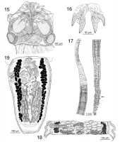
Figures 1519. Uncibilocularis squireorum n. sp. 15. Scolex. 16. Hooks. 17. Whole worm; arrow indicates location of mature proglottid drawn (Fig. 18). 18. Mature proglottid with testes. 19. Terminal p... MoreFigures 1519. Uncibilocularis squireorum n. sp. 15. Scolex. 16. Hooks. 17. Whole worm; arrow indicates location of mature proglottid drawn (Fig. 18). 18. Mature proglottid with testes. 19. Terminal proglottid. |
Line Drawing 2
|
Photo Micrograph
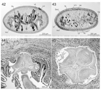
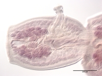
42. Uncibilocularis squireorum n. sp.; transverse section of mature proglottid with
testes at level of ovarian bridge. 43. Uncibilocularis squireorum n. sp.; transverse section of mature proglottid w... More42. Uncibilocularis squireorum n. sp.; transverse section of mature proglottid with
testes at level of ovarian bridge. 43. Uncibilocularis squireorum n. sp.; transverse section of mature proglottid with testes at
level of genital pore. 44. Uncibilocularis squireorum n. sp., longitudinal section of scolex in situ. 45. Uncibilocularis squireorum n. sp., transverse section of scolex in situ at level of anterior loculus. (Cs, cirrus sac; Ded, dorsal excretory duct; Gp, genital pore; Lm, longitudinal muscle bundle; Mg, Mehlis gland; O, ovary; T, testis; Ut, uterus; Vf, vitelline follicle; Va, vagina; Vd, vas deferens; Ved, ventral excretory duct.). |
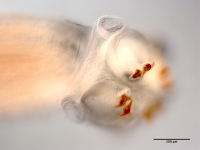
Scanning Electron Micrograph

35. U. squireorum, scolex. 36. U. squireorum, distal bothridial surface. 37. U. squireorum, proximal bothridial surface. 38. U. squireorum, surface of scolex proper. 39. U. squireorum, surface of ceph... More35. U. squireorum, scolex. 36. U. squireorum, distal bothridial surface. 37. U. squireorum, proximal bothridial surface. 38. U. squireorum, surface of scolex proper. 39. U. squireorum, surface of cephalic peduncle. (Small numbers in Fig. 35 indicate regions enlarged in corresponding figures.) |
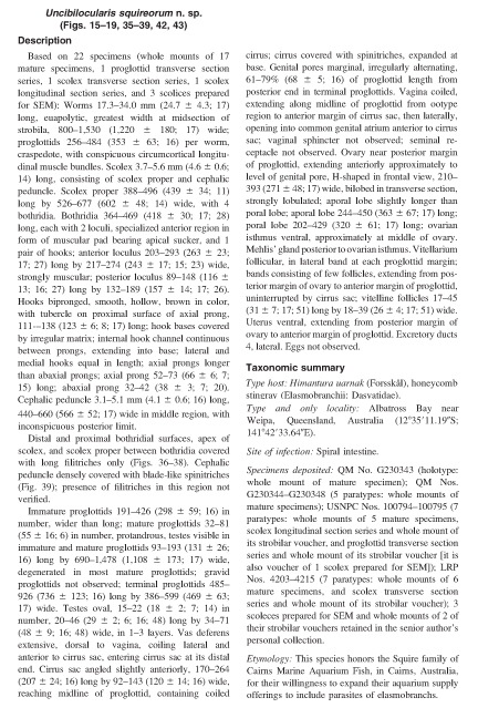
 Figures 1519. Uncibilocularis squireorum n. sp. 15. Scolex. 16. Hooks. 17. Whole worm; arrow indicates location of mature proglottid drawn (Fig. 18). 18. Mature proglottid with testes. 19. Terminal proglottid.
 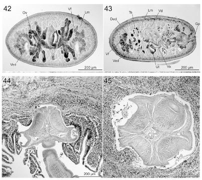 42. Uncibilocularis squireorum n. sp.; transverse section of mature proglottid with
testes at level of ovarian bridge. 43. Uncibilocularis squireorum n. sp.; transverse section of mature proglottid with testes at
level of genital pore. 44. Uncibilocularis squireorum n. sp., longitudinal section of scolex in situ. 45. Uncibilocularis squireorum n. sp., transverse section of scolex in situ at level of anterior loculus. (Cs, cirrus sac; Ded, dorsal excretory duct; Gp, genital pore; Lm, longitudinal muscle bundle; Mg, Mehlis gland; O, ovary; T, testis; Ut, uterus; Vf, vitelline follicle; Va, vagina; Vd, vas deferens; Ved, ventral excretory duct.).
42. Uncibilocularis squireorum n. sp.; transverse section of mature proglottid with
testes at level of ovarian bridge. 43. Uncibilocularis squireorum n. sp.; transverse section of mature proglottid with testes at
level of genital pore. 44. Uncibilocularis squireorum n. sp., longitudinal section of scolex in situ. 45. Uncibilocularis squireorum n. sp., transverse section of scolex in situ at level of anterior loculus. (Cs, cirrus sac; Ded, dorsal excretory duct; Gp, genital pore; Lm, longitudinal muscle bundle; Mg, Mehlis gland; O, ovary; T, testis; Ut, uterus; Vf, vitelline follicle; Va, vagina; Vd, vas deferens; Ved, ventral excretory duct.). 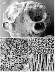 35. U. squireorum, scolex. 36. U. squireorum, distal bothridial surface. 37. U. squireorum, proximal bothridial surface. 38. U. squireorum, surface of scolex proper. 39. U. squireorum, surface of cephalic peduncle. (Small numbers in Fig. 35 indicate regions enlarged in corresponding figures.)
  QM No. G230343, holotype slide
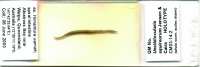
  QM G230343, scolex of holotype
  QM G230343, terminal proglottid of holotype
  USNPC No. 100794, paratype slide

