Line Drawing 1

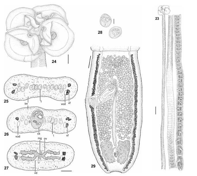
FIGURES 23. Entire worm, scale bar ! 1 mm. (23) Orygmatobothrium juani n. sp. FIGURES 2429. Orygmatobothrium juani n. sp. (24) Scolex, scale bar = 200 µm. (2527) Cross sections of mature proglottid,... MoreFIGURES 23. Entire worm, scale bar ! 1 mm. (23) Orygmatobothrium juani n. sp. FIGURES 2429. Orygmatobothrium juani n. sp. (24) Scolex, scale bar = 200 µm. (2527) Cross sections of mature proglottid, scale bar =
100 µm. (25) At level of testes anterior to cirrus sac. (26) At level of cirrus sac. (27) At level of ovarian isthmus. (28) Eggs, scale bar = 10 µm.
(29) Last mature proglottid, scale bar = 200 µm. Abbreviations: cs, cirrus sac; lm, longitudinal musculature; mg, Mehliss gland; ov, ovary; t,
testis; vd, vitelline duct; vf, vitelline follicle; vod, ventral osmoregulatory duct. |
Line Drawing 2
|
Photo Micrograph
|
Scanning Electron Micrograph
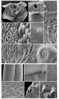
FIGURES 3038. Orygmatobothrium juani n. sp., scanning electron micrographs. (30) Scolex, scale bar = 200 µm. (31) Accessory sucker at anterior margin of bothridium, arrow indicates cleft without over... MoreFIGURES 3038. Orygmatobothrium juani n. sp., scanning electron micrographs. (30) Scolex, scale bar = 200 µm. (31) Accessory sucker at anterior margin of bothridium, arrow indicates cleft without overlapped margins, scale bar = 20 µm. (32) Detail of cephalic peduncle and germinative zone, scale bar = 50 µm. (33) Detail of microtriches on outer surface of accessory sucker, scale bar = 1 µm. (34) Distal bothridial surface, scale bar = 2 µm; inset shows enlarged detail of microtriches, scale bar = 1 µm. (35) Detail of a rounded projection with central cilium and filiform microtriches on outer surface of central glandulomuscular organ, scale bar = 1 µm. (36) Detail of microtriches on cephalic peduncle, scale bar = 2 µm. (37) Proximal bothridial surface, scale bar = 2 µm; inset shows enlarged detail of microtriches, arrows indicate position of projections in trifid microtriches, scale bar = 1 µm. (38) Glandulomuscular organ on distal bothridial surface, arrows indicate the presence of
rounded projections with cilia magnified in Figure 35, scale bar = 25 µm. |
 FIGURES 23. Entire worm, scale bar ! 1 mm. (23) Orygmatobothrium juani n. sp. FIGURES 2429. Orygmatobothrium juani n. sp. (24) Scolex, scale bar = 200 µm. (2527) Cross sections of mature proglottid, scale bar =
100 µm. (25) At level of testes anterior to cirrus sac. (26) At level of cirrus sac. (27) At level of ovarian isthmus. (28) Eggs, scale bar = 10 µm.
(29) Last mature proglottid, scale bar = 200 µm. Abbreviations: cs, cirrus sac; lm, longitudinal musculature; mg, Mehliss gland; ov, ovary; t,
testis; vd, vitelline duct; vf, vitelline follicle; vod, ventral osmoregulatory duct.
FIGURES 23. Entire worm, scale bar ! 1 mm. (23) Orygmatobothrium juani n. sp. FIGURES 2429. Orygmatobothrium juani n. sp. (24) Scolex, scale bar = 200 µm. (2527) Cross sections of mature proglottid, scale bar =
100 µm. (25) At level of testes anterior to cirrus sac. (26) At level of cirrus sac. (27) At level of ovarian isthmus. (28) Eggs, scale bar = 10 µm.
(29) Last mature proglottid, scale bar = 200 µm. Abbreviations: cs, cirrus sac; lm, longitudinal musculature; mg, Mehliss gland; ov, ovary; t,
testis; vd, vitelline duct; vf, vitelline follicle; vod, ventral osmoregulatory duct. 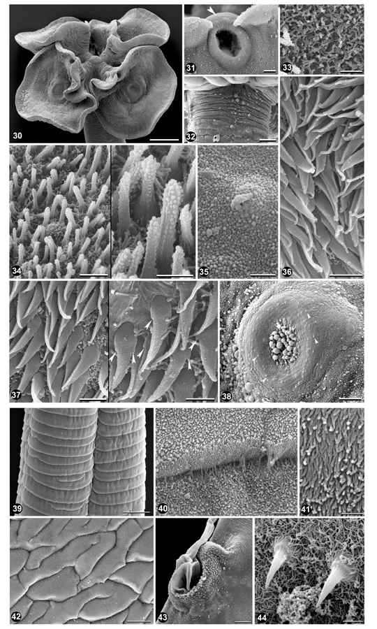 FIGURES 3038. Orygmatobothrium juani n. sp., scanning electron micrographs. (30) Scolex, scale bar = 200 µm. (31) Accessory sucker at anterior margin of bothridium, arrow indicates cleft without overlapped margins, scale bar = 20 µm. (32) Detail of cephalic peduncle and germinative zone, scale bar = 50 µm. (33) Detail of microtriches on outer surface of accessory sucker, scale bar = 1 µm. (34) Distal bothridial surface, scale bar = 2 µm; inset shows enlarged detail of microtriches, scale bar = 1 µm. (35) Detail of a rounded projection with central cilium and filiform microtriches on outer surface of central glandulomuscular organ, scale bar = 1 µm. (36) Detail of microtriches on cephalic peduncle, scale bar = 2 µm. (37) Proximal bothridial surface, scale bar = 2 µm; inset shows enlarged detail of microtriches, arrows indicate position of projections in trifid microtriches, scale bar = 1 µm. (38) Glandulomuscular organ on distal bothridial surface, arrows indicate the presence of
rounded projections with cilia magnified in Figure 35, scale bar = 25 µm.
FIGURES 3038. Orygmatobothrium juani n. sp., scanning electron micrographs. (30) Scolex, scale bar = 200 µm. (31) Accessory sucker at anterior margin of bothridium, arrow indicates cleft without overlapped margins, scale bar = 20 µm. (32) Detail of cephalic peduncle and germinative zone, scale bar = 50 µm. (33) Detail of microtriches on outer surface of accessory sucker, scale bar = 1 µm. (34) Distal bothridial surface, scale bar = 2 µm; inset shows enlarged detail of microtriches, scale bar = 1 µm. (35) Detail of a rounded projection with central cilium and filiform microtriches on outer surface of central glandulomuscular organ, scale bar = 1 µm. (36) Detail of microtriches on cephalic peduncle, scale bar = 2 µm. (37) Proximal bothridial surface, scale bar = 2 µm; inset shows enlarged detail of microtriches, arrows indicate position of projections in trifid microtriches, scale bar = 1 µm. (38) Glandulomuscular organ on distal bothridial surface, arrows indicate the presence of
rounded projections with cilia magnified in Figure 35, scale bar = 25 µm. 

