Line Drawing 1
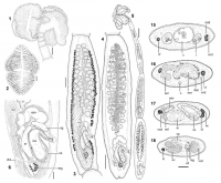
Figure 16. Rhinebothroides campbelli n. sp. 1. Scolex. 2. Detail of distal surface of bothridium. 3. Mature segment, dorsal view. 4. Gravid
segment, ventral view. 5. Entire worm. 6. Detail of genita... MoreFigure 16. Rhinebothroides campbelli n. sp. 1. Scolex. 2. Detail of distal surface of bothridium. 3. Mature segment, dorsal view. 4. Gravid
segment, ventral view. 5. Entire worm. 6. Detail of genitalia. Abbreviations: cs, cirrus-sac; esv, external seminal vesicle; isv, internal seminal
vesicle; mg, Mehlis gland; ov, ovary; sr, seminal receptacle; u, uterus; vg, vagina; vtd, vitelline duct. Scale-bars: 1,3, 100 μm; 2,6, 50 μm; 4, 200 μm; 5, 300 μm. Figure 1518. Rhinebothroides campbelli n. sp., cross-sections of gravid segment. 15. At level of testes. 16. Slightly anterior to the genital pore. 17. At level of cirrus-sac below the genital pore. 18. At level of Mehlis gland. Abbreviations: aol, aporal ovarian lobe; cs, cirrus-sac; dod, dorsal osmoregulatory duct; dsc, darkly staining
cells sourrending external seminal vesicle; esv, external seminal vesicle; mg, Mehlis gland; ov, ovary; nc, nerve cord; sr, seminal receptacle; t, testes; u, uterus; vf, vitelline follicles; vg, vagina; vod, ventral osmoregulatory duct. Scale-bar: 50 μm |
Line Drawing 2
|
Photo Micrograph
|
Scanning Electron Micrograph
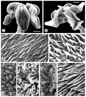
Figure 714. Rhinebothroides campbelli n. sp., scanning electron micrographs. 7-8. Scolex. 9. Proximal bothridial surface. 10. Distal bothridial
surface. 11. Stalk surface. 12. Germinative zone surfa... MoreFigure 714. Rhinebothroides campbelli n. sp., scanning electron micrographs. 7-8. Scolex. 9. Proximal bothridial surface. 10. Distal bothridial
surface. 11. Stalk surface. 12. Germinative zone surface. 13. Surface of gravid segment near anterior margin. 14. Surface of gravid segment near posterior margin. Scale-bars: 7,8, 100 μm; 9,14, 2 μm; 10-13, 1 μm. |


 Figure 16. Rhinebothroides campbelli n. sp. 1. Scolex. 2. Detail of distal surface of bothridium. 3. Mature segment, dorsal view. 4. Gravid
segment, ventral view. 5. Entire worm. 6. Detail of genitalia. Abbreviations: cs, cirrus-sac; esv, external seminal vesicle; isv, internal seminal
vesicle; mg, Mehlis gland; ov, ovary; sr, seminal receptacle; u, uterus; vg, vagina; vtd, vitelline duct. Scale-bars: 1,3, 100 μm; 2,6, 50 μm; 4, 200 μm; 5, 300 μm. Figure 1518. Rhinebothroides campbelli n. sp., cross-sections of gravid segment. 15. At level of testes. 16. Slightly anterior to the genital pore. 17. At level of cirrus-sac below the genital pore. 18. At level of Mehlis gland. Abbreviations: aol, aporal ovarian lobe; cs, cirrus-sac; dod, dorsal osmoregulatory duct; dsc, darkly staining
cells sourrending external seminal vesicle; esv, external seminal vesicle; mg, Mehlis gland; ov, ovary; nc, nerve cord; sr, seminal receptacle; t, testes; u, uterus; vf, vitelline follicles; vg, vagina; vod, ventral osmoregulatory duct. Scale-bar: 50 μm
Figure 16. Rhinebothroides campbelli n. sp. 1. Scolex. 2. Detail of distal surface of bothridium. 3. Mature segment, dorsal view. 4. Gravid
segment, ventral view. 5. Entire worm. 6. Detail of genitalia. Abbreviations: cs, cirrus-sac; esv, external seminal vesicle; isv, internal seminal
vesicle; mg, Mehlis gland; ov, ovary; sr, seminal receptacle; u, uterus; vg, vagina; vtd, vitelline duct. Scale-bars: 1,3, 100 μm; 2,6, 50 μm; 4, 200 μm; 5, 300 μm. Figure 1518. Rhinebothroides campbelli n. sp., cross-sections of gravid segment. 15. At level of testes. 16. Slightly anterior to the genital pore. 17. At level of cirrus-sac below the genital pore. 18. At level of Mehlis gland. Abbreviations: aol, aporal ovarian lobe; cs, cirrus-sac; dod, dorsal osmoregulatory duct; dsc, darkly staining
cells sourrending external seminal vesicle; esv, external seminal vesicle; mg, Mehlis gland; ov, ovary; nc, nerve cord; sr, seminal receptacle; t, testes; u, uterus; vf, vitelline follicles; vg, vagina; vod, ventral osmoregulatory duct. Scale-bar: 50 μm  Figure 714. Rhinebothroides campbelli n. sp., scanning electron micrographs. 7-8. Scolex. 9. Proximal bothridial surface. 10. Distal bothridial
surface. 11. Stalk surface. 12. Germinative zone surface. 13. Surface of gravid segment near anterior margin. 14. Surface of gravid segment near posterior margin. Scale-bars: 7,8, 100 μm; 9,14, 2 μm; 10-13, 1 μm.
Figure 714. Rhinebothroides campbelli n. sp., scanning electron micrographs. 7-8. Scolex. 9. Proximal bothridial surface. 10. Distal bothridial
surface. 11. Stalk surface. 12. Germinative zone surface. 13. Surface of gravid segment near anterior margin. 14. Surface of gravid segment near posterior margin. Scale-bars: 7,8, 100 μm; 9,14, 2 μm; 10-13, 1 μm. 

