Cestode Scientific Name
| Species ID | 5583 |
|---|---|
| Order | Tetrabothriidea |
| Family | |
| Subfamily | |
| Genus | Tetrabothrius (Tetrabothrius) |
| Species | shinni |
| Authority | Hoberg, 1987 |
| Taxonomic Status | Valid |
| Valid Name | |
| Synonyms | |
| Genus Record | No |
| Type Species | |
| Verified | No |
| Verified By | |
| Citation(s) |
Hoberg, E. P. 1987. Tetrabothrius shinni sp.n. (Eucestoda), from Phalacrocorax atriceps bransfieldensis (Pelecaniformes) in Antarcita, with comments on morphological variation and host-parasite biogeography and evolution. Canadian Journal of Zoology 65: 2969-2975. (4218) Download PDF |
| Redescription | |
| Scientific Name Notes |
Record Data
| Date (MM/DD/YYYY) | Action | User Name |
|---|---|---|
| 11/30/-0001 | Created | K. Catanese |
| 02/18/2010 | Modified | |
| 02/11/2016 | Modified | K. Mojica |
Type Host
| Host Class | |||||||
|---|---|---|---|---|---|---|---|
| Host Order | |||||||
| Host Family | |||||||
|
Type Host (Literal) |
|
||||||
|
Type Host (Valid) |
|
||||||
| Additional Host(s) | |||||||
| Site in Host | middle 1/3 of small intestine | ||||||
| Host Notes |
Type Locality
| Country | Antarctica |
|---|---|
| Body of Water | |
| Island(s) | |
| City/Region | Cormorant Island, Arthur Harbor, Anvers Island |
| Coordinates | |
| DD Latitude | ca. 64°46S |
| DD Longitude | 64°05W |
| Additional Localities | |
| Locality Notes |
Specimens
| Type Material | USNM Helm. Coll. No. 79657 (holotype) USNM No. 79658 (paratype) |
|---|---|
| Total Number of Type Specimens | |
| Voucher Material | No. 79659 (two voucher specimens) |
| Specimen Notes |
Data are given as in original description unless otherwise indicated.
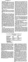
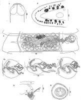
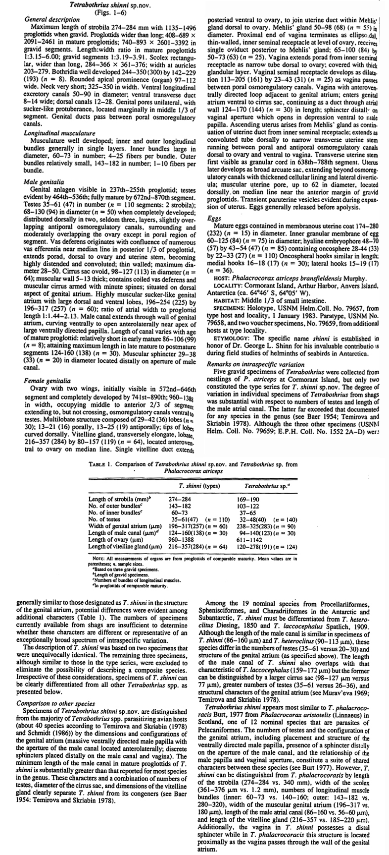
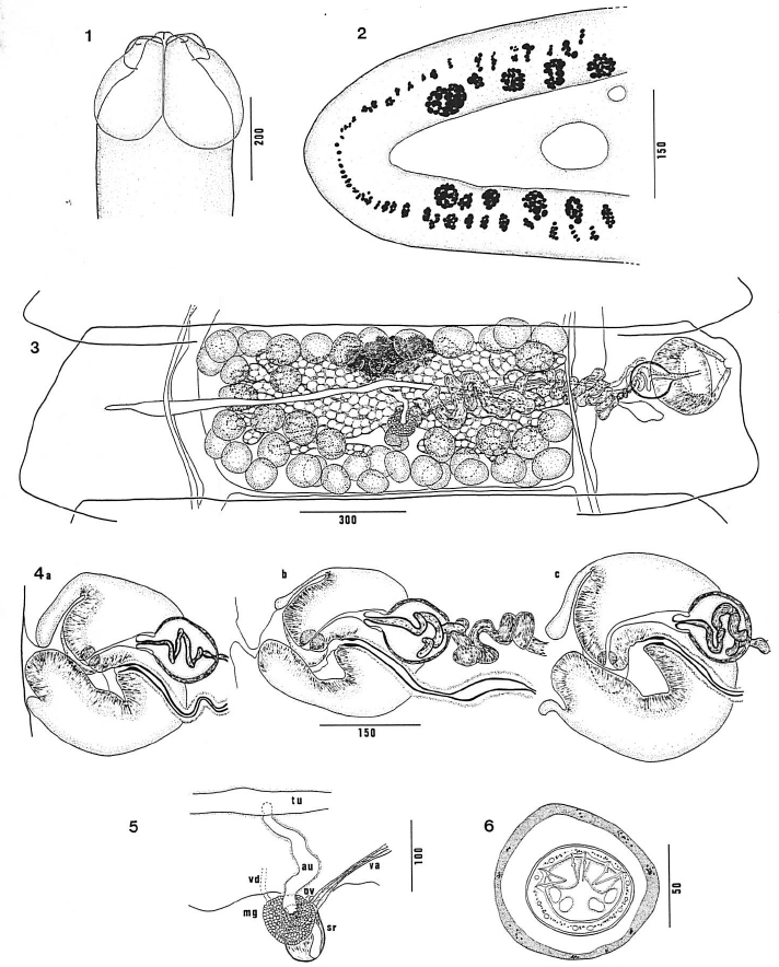 FIGS. 1-6. Tetrabothrius shinn; sp. nov. Fig. 1. Scolex. Fig. 2. Distribution of longitudinal muscle bundles in antiporal portion of a mature proglottid, transverse section. Fig. 3. Mature proglottid, dorsal view. Fig. 4. Detail of genital atrium showing male and female duels, and cirrus sac as viewed from the anterior in transverse section. Note change in length of male canal in relation to maturity in a single specimen: (a) early mature; (b) mature; also showing detail of vaginal seminal receptacle; (c) postmature. Fig. 5. Detail of female genital ducts, dorsal view, showing vagina (va), inner seminal receptacle (ST), oviduct (ov) , Mehlis' gland (mg) . vitelline duct (vel). ascending uterine duct (au), and transverse utenne stem (tu). Fig. 6. Egg. showing inner granular membrane, hyaline embryophore, and hexacanth embryo. All scale bars are in micrometres.
 Best viewed in Firefox


