Cestode Scientific Name
| Species ID | 4858 |
|---|---|
| Order | Trypanorhyncha |
| Family | Lacistorhynchidae |
| Subfamily | |
| Genus | Pseudogrillotia |
| Species | epinepheli |
| Authority | (Scholz, Garippa & Scala, 1993) Palm, 2004 |
| Taxonomic Status | Valid |
| Valid Name | |
| Synonyms | Grillotia (Progrillotia) epinepheli Scholz, Garippa & Scala, 1993 |
| Genus Record | No |
| Type Species | No |
| Verified | Yes |
| Verified By | I. Beveridge |
| Citation(s) |
Scholz, T., G. Garippa and A. Scala. 1993. Grillotia epinepheli sp. n. (Cestoda: Trypanorhyncha) plerocerci from the teleost, Epinepheleus guaza, in Sardinia, Italy. Folia Parasitologica 40: 23-28. (4364) Download PDFPalm, H. W. 2004. The Trypanorhyncha Diesing, 1863. PKSPL-IPB Press, Bogor, 710 pp. (4253) Download PDF |
| Redescription | |
| Scientific Name Notes |
Record Data
| Date (MM/DD/YYYY) | Action | User Name |
|---|---|---|
| 11/30/-0001 | Created | I. Beveridge |
| 05/07/2015 | Modified | |
| 05/24/2015 | Modified | I. Beveridge |
| 08/16/2017 | Modified | K. Herzog |
Type Host
| Host Class | Actinopterygii | ||||||
|---|---|---|---|---|---|---|---|
| Host Order | Perciformes | ||||||
| Host Family | Serranidae | ||||||
|
Type Host (Literal) |
|
||||||
|
Type Host (Valid) |
|
||||||
| Additional Host(s) | |||||||
| Site in Host | peritoneum, surface of internal organs | ||||||
| Host Notes |
Type Locality
| Country | Italy |
|---|---|
| Body of Water | Mediterranean Sea |
| Island(s) | |
| City/Region | Isola Molara near Porto San Paolo, Sardinia |
| Coordinates | 40°52'N, 9°85E |
| DD Latitude | |
| DD Longitude | |
| Additional Localities | |
| Locality Notes |
Specimens
| Type Material | IPCAS C-221 (holotype, paratypes & specimens on SEM stubs); BMNH 1992.10.6.1-2 (paratypes); USNPC 82612 (paratypes); Cattedra Malattie Parassitarie, Universita di Sassari, Italia (paratypes) |
|---|---|
| Total Number of Type Specimens | 1 holotype, 8 paratypes, and an unknown number of specimens prepared for SEM |
| Voucher Material | |
| Specimen Notes |
Data are given as in original description unless otherwise indicated.

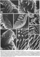
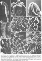
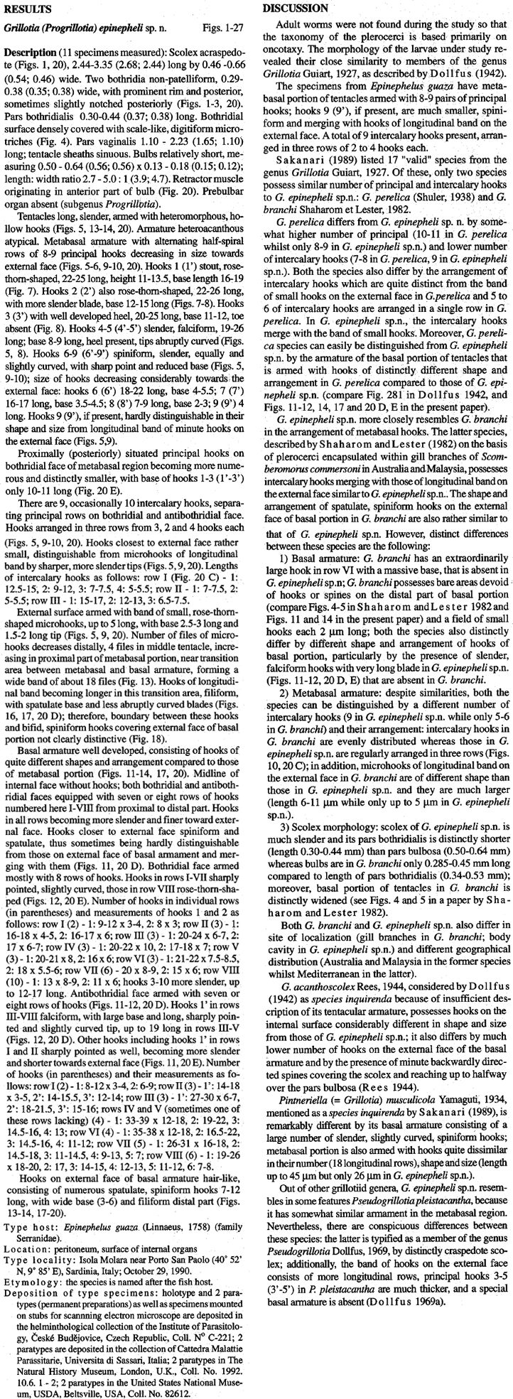
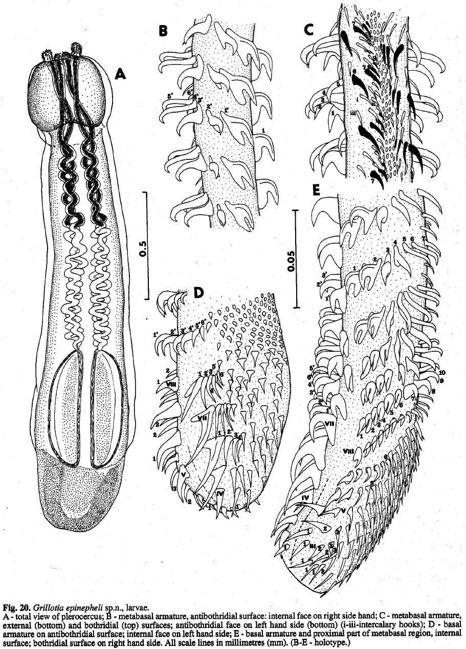 Fig. 20. Grillotia epinepheli sp.n., larvae. A - total view of plerocercus;B -metabasala rmature,a ntibothridial surface: internal face on right side hand; C -metabasal armature, external (bottom) and bothridial (top) surfaces; antibothridialf ace on left hand side (bottom) (i-iii-intercalaryh ooks); D - basal
armature on antibothridial surface; internal face on left hand side; E - basal armature and proximal part of metabasal region, internal surface; bothridial surface on right hand side. All scale lines in millimetres (mm). (B-E - holotype.)
Fig. 20. Grillotia epinepheli sp.n., larvae. A - total view of plerocercus;B -metabasala rmature,a ntibothridial surface: internal face on right side hand; C -metabasal armature, external (bottom) and bothridial (top) surfaces; antibothridialf ace on left hand side (bottom) (i-iii-intercalaryh ooks); D - basal
armature on antibothridial surface; internal face on left hand side; E - basal armature and proximal part of metabasal region, internal surface; bothridial surface on right hand side. All scale lines in millimetres (mm). (B-E - holotype.) 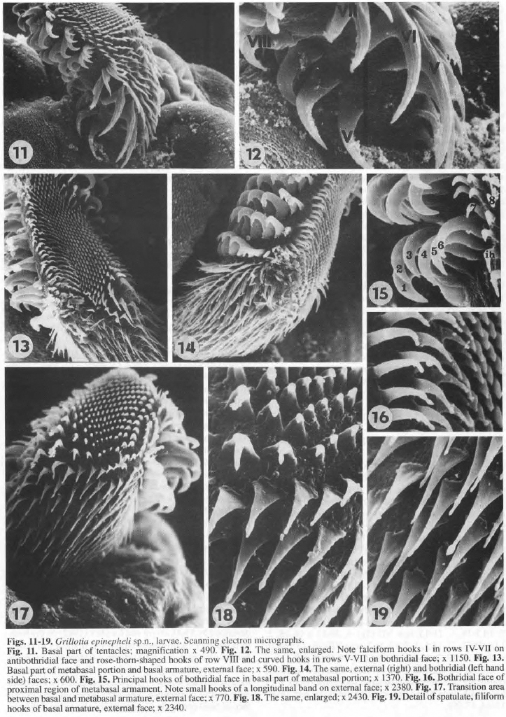 Figs. 11-19. Grillotia epinepheli sp.n., larvae. Scanning electron micrographs. Fig. 11. Basal part of tentacles; magnification x 490. Fig. 12. The same, enlarged. Note falcifonn hooks I in rows IV-VU on antibothridial face and rose-thorn-shaped hooks of row YUi and curved hooks in rows V-VII on bothridial face; x 1150. Fig. 13. Basal part of metabasal portion and basal armature , externa l face; x 590. Fig. 14. The same, external (right) and bothridial (left hand side) faces; x 600. Fig. 15. Principal hooks of bothridial face in basal part of metabasal portion; x 1370. Fig. 16. Bothridial face of proximal region of metabasal annament. Note small hooks of a longitudinal band on external face; x 2380. Fig. 17. Transition area between basal and metabasal armature , external face; x 770. Fig.18. The same, enlarged; x 2430. Fig. 19. Detail of spatulate, filiform hooks of basal armature, external face; x 2340.
  Figs. 1-10. Grillotia epinepheli sp.n., larvae. Scanning electron micrographs. Fig. 1. Total view of larva; magnification x 40. Fig. 2. Scolex with partly everted tentacles; x 192. Fig. 3. Scolex with completely withdrawn tentacles; x 170. Fig. 4. Detail of scale-like, digitiform tegumental microtriches cover ing bothridia; x 1480. Fig. 5. Metabasal portion with longitudinal band of sma ll hooks on external face, and intercalary and principal hooks on margins; x 720. Fig. 6. lntercalary (ih) and principal hooks of metabasal armature, antibothridial face; x 1380. Fig. 7. Detail of principal hooks 1-2 of metabasal armature; x 950. Fig. 8. Principal hooks of metabasal armature; x 750. Fig. 9. Detail of external face of melabasal portion; note three rows of intercalary hooks (i:l-Ill) and small principal hooks 9 (arrows); x 940. Fig. 10. Detail of principal hooks 3-8 (hook 9 not present) and intercalary hooks (i) arranged in three rows (I-Ill ); x 24 10.
 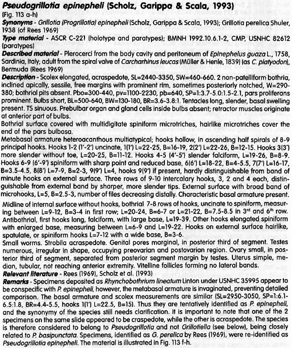 Palm (2004), pg. 265
 Best viewed in Firefox


