Cestode Scientific Name
| Species ID | 4855 |
|---|---|
| Order | Trypanorhyncha |
| Family | Lacistorhynchidae |
| Subfamily | |
| Genus | Grillotia |
| Species | borealis |
| Authority | Keeney & Campbell, 2001 |
| Taxonomic Status | Valid |
| Valid Name | |
| Synonyms | |
| Genus Record | No |
| Type Species | No |
| Verified | Yes |
| Verified By | I. Beveridge |
| Citation(s) |
Keeney, D. B. and R. A. Campbell. 2001. Grillotia borealis sp. n. (Cestoda: Trypanorhyncha) from five species of Bathyraja (Rajiiformes: Arhynchobatidae) in the North Pacific Ocean with comments on parasite enteric distribution. Folia Parasitologica 48: 21-29. (4362) Download PDF |
| Redescription | |
| Scientific Name Notes | Redescribed by Beveridge & Campbell (2007) (4523) |
Record Data
| Date (MM/DD/YYYY) | Action | User Name |
|---|---|---|
| 11/30/-0001 | Created | I. Beveridge |
| 01/12/2010 | Modified | |
| 05/21/2015 | Modified | I. Beveridge |
| 05/29/2017 | Modified | K. Herzog |
| 06/06/2017 | Modified | K. Herzog |
| 07/27/2017 | Modified | K. Jensen |
Type Host
| Host Class | Elasmobranchii | ||||||
|---|---|---|---|---|---|---|---|
| Host Order | Rajiformes | ||||||
| Host Family | Arhynchobatidae | ||||||
|
Type Host (Literal) |
|
||||||
|
Type Host (Valid) |
|
||||||
| Additional Host(s) | Bathyraja aleutica; Bathyraja interrupta; Bathyraja minispinosa; Bathyraja smirnova | ||||||
| Site in Host | spiral intestine | ||||||
| Host Notes |
Type Locality
| Country | |
|---|---|
| Body of Water | Bering Sea |
| Island(s) | |
| City/Region | |
| Coordinates | 61°0053N, 173°2691WW |
| DD Latitude | |
| DD Longitude | |
| Additional Localities | Bering Sea: (56°5988N, 167°4175W); (56°5047N, 170°2859W); Sea of Okhotsk (coordinates not available) |
| Locality Notes |
Specimens
| Type Material | USNPC 90678 (holotype), USNPC 90679-81, IPCAS C-336 (paratypes) |
|---|---|
| Total Number of Type Specimens | |
| Voucher Material | see Keeney & Campbell (2001) for relation of voucher material |
| Specimen Notes |
Data are given as in original description unless otherwise indicated.
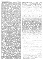
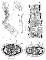


 Figs. 1-6. Grillotia borealis sp. n. Fig. 1. Scolex. Fig. 2. Pars bothridialis. Fig. 3. Bulb. Fig. 4. Mature segment, vitelline follicles only shown on lateral margins for clarity. Fig. 5. Mature segment, cross-section anterior to genital pore. Fig. 6. Mature segment, cross-section through ovary. Abbreviations: DOC - dorsal osmoregulatory canal; M - longitudinal muscle; OV - ovary; T - testis; U - uterus; VOC - ventral osmoregulatory canal; VT - vitelline follicle. Scale bars: Fig. 1 = 700 µm; Figs. 2, 3 = 50 µm; Fig. 4 = 300 µm; Figs. 5, 6 = 200 µm.
 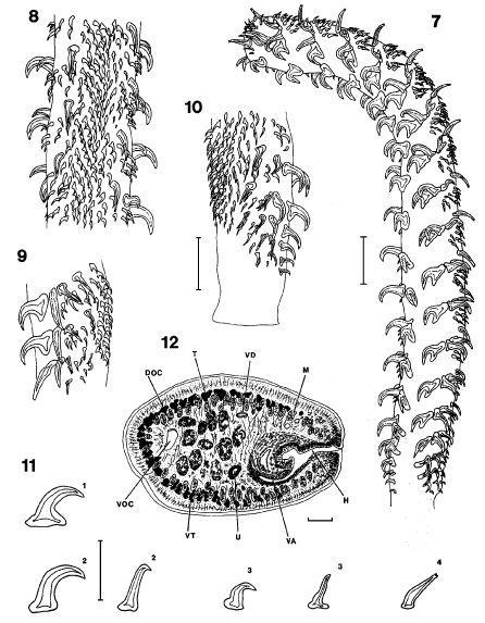 Figs. 7-12. Grillotia borealis sp. n. Fig. 7. Tentacle, internal surface at left. Fig. 8. Band of microhooks on external surface of metabasal region of tentacle. Fig. 9. Metabasal armature, bothridial surface. Fig. 10. Basal armature, bothridial surface; internal surface at right. Fig. 11. Profiles of principal hook, note change in form of hooks 2(2') and 3(3'). Fig. 12. Cross-section through hermaphroditic sac, note junction of vagina with male duct in hermaphroditic sac. Abbreviations: DOC - dorsal osmoregulatory canal; H - hermaphroditic duct; M - longitudinal muscle; OV - ovary; T - testis; U - uterus; VA - vagina; VD - vas deferens; VOC - ventral osmoregulatory canal; VT - vitelline follicle. Scale bars: Fig. 7 = 100 µm; Figs. 8-10 = 500 µm; Fig. 11 = 50 µm; Fig. 12 = 20 µm.
 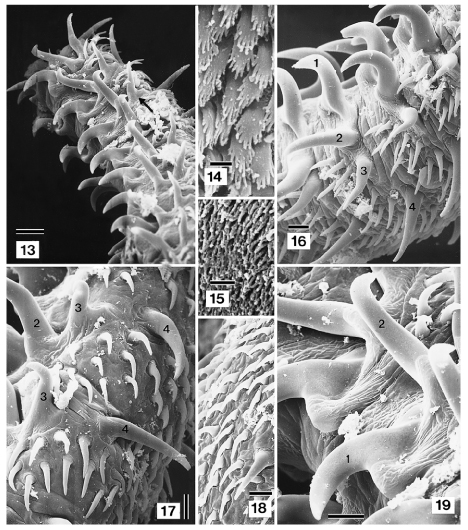 Figs. 13-19. Grillotia borealis sp. n., scanning electron micrographs. Fig. 13. Distal metabasal armature. Note crochet-hook shape of hooks 2(2'). The transverse orientation of the bases of principal hooks 3 is clearly visible (arrow). Fig. 14. Pectinate microtriches on surface of bothridium. Fig. 15. Filiform microtriches on surface of pars bulbosa. Fig. 16. Metabasal armature, bothridial surface, principal hooks numbered (1-4). Fig. 17. Metabasal armature. Note change in morphology of hooks 3, notched tips of hook 4, and intercalary rows. Fig. 18. Proximal metabasal armature. Note merging of intercalary rows with band of microhooks on external surface at left. Fig. 19. Detail of hook files 1 and 2 in upper metabasal region. Scale bars: Figs. 13, 16-19 = 10 µm; Figs. 14, 15 = 1 µm.
 Best viewed in Firefox


