Cestode Scientific Name
| Species ID | 4853 |
|---|---|
| Order | Trypanorhyncha |
| Family | Lacistorhynchidae |
| Subfamily | |
| Genus | Rhynchobothrium |
| Species | ingens |
| Authority | Linton, 1921 |
| Taxonomic Status | Synonym |
| Valid Name | Floriceps saccatus Cuvier, 1817 |
| Synonyms | |
| Genus Record | No |
| Type Species | No |
| Verified | Yes |
| Verified By | I. Beveridge |
| Citation(s) |
Linton, E. 1921. Rhynchobothrium ingens spec. nov. a parasite of the dusky shark (Carcharhinus obscurus). Journal of Parasitology 8(1): 22-32. (279) Download PDF |
| Redescription | |
| Scientific Name Notes |
Record Data
| Date (MM/DD/YYYY) | Action | User Name |
|---|---|---|
| 11/30/-0001 | Created | I. Beveridge |
| 03/24/2010 | Modified | |
| 05/24/2015 | Modified | I. Beveridge |
| 05/17/2016 | Modified | B. Barbeau |
Type Host
| Host Class | |||||||
|---|---|---|---|---|---|---|---|
| Host Order | |||||||
| Host Family | |||||||
|
Type Host (Literal) |
|
||||||
|
Type Host (Valid) |
|
||||||
| Additional Host(s) | |||||||
| Site in Host | spiral valve | ||||||
| Host Notes |
Type Locality
| Country | U.S.A. |
|---|---|
| Body of Water | Atlantic Ocean |
| Island(s) | |
| City/Region | Menemsha La Bight |
| Coordinates | |
| DD Latitude | |
| DD Longitude | |
| Additional Localities | |
| Locality Notes |
Specimens
| Type Material | USNPC 88568 (type) |
|---|---|
| Total Number of Type Specimens | |
| Voucher Material | |
| Specimen Notes |
Data are given as in original description unless otherwise indicated.

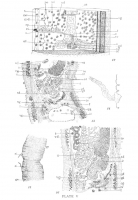

 EXPLANATION OF PLATE IV: Plate IV.-Fig. 1. - Scolex of Rhynchobothrium ingens. Diagrammatic sketch of scolex, marginal view, showing distribution of ganglion cells; length of scolex 9.5 mm. Fig. 2. - Transverse section of bothria, showing anastomosing excretory vessels, retracted proboscides, etc. Dorso-ventral diameter 1.16 mm. Fig. 3. - Enlarged view of section of border of Bothrium. Fig. 4. - Transverse section of neck of scolex showing ganglion cells, excretory vessels, etc. Shorter diameter of section 1 mm. Fig. 5. - Diagram of proboscis, split longitudinally and partly flattened; for significance of letters a-e, see text. Fig. 6. - Bothrium, front view; breadth 2 mm. Fig. 7. - Two of larger hooks of series b, Fig. 5, seen in side view, optical section. Fig. 8.-Hook from series e, Figure 5. Fig. 9. - Hooks from series b, Figure 5. Fig. 10. - Hook from series d, Figure 5. Fig. 11. - Median region of transverse section made near the posterior end of a mature proglottis, dorso-ventral diameter 1 mm.
  EXPLANATION OF PLATE V: Plate V.-Fig. 12. - Stereogram of mature proglottis, ventral view. Fig. 13. - Median sagittal section at junction of two mature proglottides; dorso-ventral diameter 1 mm. Three adjacent sections were used in order to show the sperm duct and vitelline duct joining the germ duct. Fig. 14. - Enlarged view of everted cirrus, length 2.3 m.m. Fig. 15. - Portion of strobile representing four proglottides. Note that the genital aperture is visible in the lower, while it is not visible in the upper proglottis. This is because the apertures are not quite symmetrically placed on the margins of the proglottides; breadth 5.5 mm. Fig. 16. - Schematic sagittal view of reproductive organs of adult proglottis in median region of proglottis. For explanation of lettering on all figures see p. 31.
 Best viewed in Firefox


