Cestode Scientific Name
| Species ID | 4840 |
|---|---|
| Order | Trypanorhyncha |
| Family | Lacistorhynchidae |
| Subfamily | |
| Genus | Grillotia |
| Species | musculara |
| Authority | (Hart, 1936) Dollfus, 1942 |
| Taxonomic Status | Valid |
| Valid Name | |
| Synonyms | Tentacularia musculara Hart, 1936 |
| Genus Record | No |
| Type Species | No |
| Verified | Yes |
| Verified By | I. Beveridge |
| Citation(s) |
Hart, J. F. 1936. Cestodes from fishes of Puget Sound - II. Tetrarhynchoidea. Transactions of the American Microscopical Society 55: 369-387. (4349) Download PDFDollfus, R.-P. 1942. Études critiques sur les tétrarhynques du Muséum de Paris. Archives du Muséum National dHistoire Naturelle, 6. Série 19: 7-466. (170) Download PDF |
| Redescription |
Beveridge, I. and R. A. Campbell. 2007. Revision of the Grillotia erinaceus (van Beneden, 1858) species complex (Cestoda: Trypanorhyncha), with the description of G. brayi n. sp.. Systematic Parasitology 68: 1-31. (4523) Download PDF |
| Scientific Name Notes | Also redescribed by Dollfus (1942) when moved into new combination (pg. 375). |
Record Data
| Date (MM/DD/YYYY) | Action | User Name |
|---|---|---|
| 11/30/-0001 | Created | I. Beveridge |
| 05/07/2015 | Modified | |
| 05/21/2015 | Modified | I. Beveridge |
| 05/11/2016 | Modified | B. Barbeau |
| 05/29/2017 | Modified | K. Herzog |
| 06/07/2017 | Modified | K. Herzog |
| 08/03/2020 | Modified | P. Lopez Dineen |
| 09/07/2022 | Modified | V. Bueno |
Type Host
| Host Class | Elasmobranchii | ||||||
|---|---|---|---|---|---|---|---|
| Host Order | Rajiformes | ||||||
| Host Family | Rajidae | ||||||
|
Type Host (Literal) |
|
||||||
|
Type Host (Valid) |
|
||||||
| Additional Host(s) | |||||||
| Site in Host | spiral intestine | ||||||
| Host Notes | see redescription in Beveridge & Campbell (2007) for additional hosts |
Type Locality
| Country | U.S.A. |
|---|---|
| Body of Water | Puget Sound |
| Island(s) | |
| City/Region | Washington |
| Coordinates | |
| DD Latitude | |
| DD Longitude | |
| Additional Localities | |
| Locality Notes | see redescription in Beveridge & Campbell (2007) for additional localities |
Specimens
| Type Material | Holotype, coll. J.F. Hart in MNHN (missing) (as per Beveridge & Campbell, 2007) |
|---|---|
| Total Number of Type Specimens | |
| Voucher Material | |
| Specimen Notes | see redescription in Beveridge & Campbell (2007) for material examined for redescription |
Data are given as in original description unless otherwise indicated.

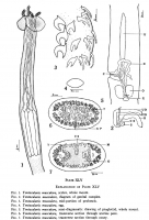

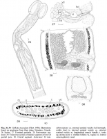
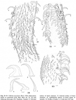
 FIG. 1. Tentacularia musculara, scolex, whole mount.
FIG. 2. Tentacularia musculara, diagram of genital complex.
FIG. 3. Tentacularia musculara, mid-portion of proboscis.
FIG. 4. Tentacularia musculara, egg.
FIG. 5. Tentacularia musculara, semi-diagramatic drawing of proglottid, whole mount.
FIG. 6. Tentacularia musculara, transverse section through uterine pore.
FIG. 7. Tentacularia musculara, transverse section through ovary.
FIG. 1. Tentacularia musculara, scolex, whole mount.
FIG. 2. Tentacularia musculara, diagram of genital complex.
FIG. 3. Tentacularia musculara, mid-portion of proboscis.
FIG. 4. Tentacularia musculara, egg.
FIG. 5. Tentacularia musculara, semi-diagramatic drawing of proglottid, whole mount.
FIG. 6. Tentacularia musculara, transverse section through uterine pore.
FIG. 7. Tentacularia musculara, transverse section through ovary. 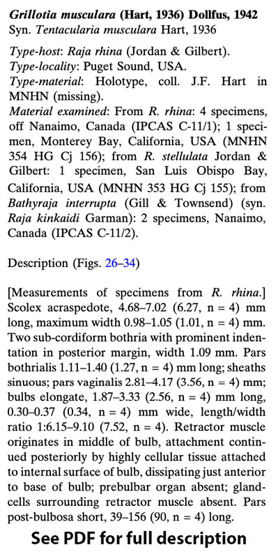
 Figs. 2630 Grillotia musculara (Hart, 1936), illustrations
based on specimens from Raja rhina, Nanaimo, Canada.
26. Scolex. 27. Terminal genitalia. 28. Post-mature segment.
29. Transverse section of mature segment at level of
genital pore. 30. Gravid segment. Scale-bars: 0.1 mm. Abbreviations: es, external seminal vesicle; hd, hermaphroditic
duct; is, internal seminal vesicle; es, external
seminal vesicle; m, longitudinal muscle bundle; t, testis
up, uterine pore; v, vitelline follicle; va, vagina; vo, ventral
osmoregulatory canal
Figs. 2630 Grillotia musculara (Hart, 1936), illustrations
based on specimens from Raja rhina, Nanaimo, Canada.
26. Scolex. 27. Terminal genitalia. 28. Post-mature segment.
29. Transverse section of mature segment at level of
genital pore. 30. Gravid segment. Scale-bars: 0.1 mm. Abbreviations: es, external seminal vesicle; hd, hermaphroditic
duct; is, internal seminal vesicle; es, external
seminal vesicle; m, longitudinal muscle bundle; t, testis
up, uterine pore; v, vitelline follicle; va, vagina; vo, ventral
osmoregulatory canal  Figs. 3134 Grillotia musculara (Hart, 1936), illustrations
based on specimens from Raja rhina (31, 33, 34) and
Bathyraja interrupta (32), Nanaimo, Canada. 31. External
surface of basal armature. 32. Internal surface of basal
armature. 33. Antibothrial surface of basal region of
tentacle. 34. Profiles of hooks 14. Scale-bars: 0.01 mm
Figs. 3134 Grillotia musculara (Hart, 1936), illustrations
based on specimens from Raja rhina (31, 33, 34) and
Bathyraja interrupta (32), Nanaimo, Canada. 31. External
surface of basal armature. 32. Internal surface of basal
armature. 33. Antibothrial surface of basal region of
tentacle. 34. Profiles of hooks 14. Scale-bars: 0.01 mm Best viewed in Firefox


