Cestode Scientific Name
| Species ID | 4837 |
|---|---|
| Order | Trypanorhyncha |
| Family | Lacistorhynchidae |
| Subfamily | |
| Genus | Floriceps |
| Species | minacanthus |
| Authority | Campbell & Beveridge, 1987 |
| Taxonomic Status | Valid |
| Valid Name | |
| Synonyms | |
| Genus Record | No |
| Type Species | No |
| Verified | Yes |
| Verified By | I. Beveridge |
| Citation(s) |
Campbell, R. A. and I. Beveridge. 1987. Floriceps minacanthus sp. nov. (Cestoda: Trypanorhyncha) from Australian fishes. Transactions of the Royal Society of South Australia 111(4): 189-194. (4348) Download PDF |
| Redescription |
Richmond, C. and J.N. Caira. 1991. Morphological investigations into Floriceps minacanthus (Trypanorhyncha: Lacistorhynchidae) with analysis of the systematic utility of scolex microtriches.. Systematic Parasitology 19: 25-32. (6533) Download PDF |
| Scientific Name Notes |
Record Data
| Date (MM/DD/YYYY) | Action | User Name |
|---|---|---|
| 11/30/-0001 | Created | I. Beveridge |
| 06/03/2014 | Modified | |
| 05/28/2015 | Modified | I. Beveridge |
| 05/29/2017 | Modified | K. Herzog |
| 06/05/2017 | Modified | K. Herzog |
| 06/06/2017 | Modified | K. Herzog |
| 07/27/2017 | Modified | K. Jensen |
Type Host
| Host Class | Elasmobranchii | ||||||
|---|---|---|---|---|---|---|---|
| Host Order | Carcharhiniformes | ||||||
| Host Family | Carcharhinidae | ||||||
|
Type Host (Literal) |
|
||||||
|
Type Host (Valid) |
|
||||||
| Additional Host(s) | Adult: Carcharhinus amboinensis; Plerocerci: Plectropomus leopardus, Platycephalus laevigatus, Platycephalus sp., Sphyraena novaehollandiae | ||||||
| Site in Host | spiral intestine | ||||||
| Host Notes |
Type Locality
| Country | Australia |
|---|---|
| Body of Water | Pacific Ocean |
| Island(s) | |
| City/Region | Tathra, New South Wales |
| Coordinates | |
| DD Latitude | |
| DD Longitude | |
| Additional Localities | Port Lincoln, South Australia; S. Lawrence, Queensland, Australia; Heron Islands, Queensland, Australia; Northhaven, South Australia; Adelaide, South Australia; |
| Locality Notes |
Specimens
| Type Material | SAM V4035 (holotype), SAM V4036-7, S2650-1, USNPC 79545-6 (paratypes) |
|---|---|
| Total Number of Type Specimens | Measurements of eight adult specimens from Carcharhinus brachyurus (types). |
| Voucher Material | |
| Specimen Notes |
Data are given as in original description unless otherwise indicated.

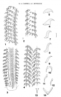
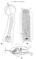
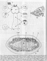
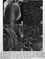
 Fig 1-14. Floriceps minacanthus sp. nov. 1,2, metabasal armature, bothridial surface; 3, basal and metabasal armature of tentacle, external surface; 4, metabasal armature, internal surface; 5-11, profiles of hooks 1, 2, 3, 4, 5, 6 and 7 respectively; 12, profile of satellite hook or hook 8; 13, chainette element from mid-tentacular region; 14, chainette element from metabasal region. Scale lines: figs 1-4, 0.1 mm; figs 5-14, 0.01 mm. Legend: bothridial hooks 1, 2 . . . . . 7; antibothridial hooks 1', 2' . . . . . 7'; satellite hooks S; chainette C.
  Figs 15-17. Floriceps minacanthus sp. nov. 15, scolex; 16, maure proglottis; 17, cirrus sac and distal vagina. Scale lines: figs 15, 16, 1.0 mm; fig. 17, 0.1 mm.
 Figs 1-3. Floriceps minacanthus. Schematic diagram summarising microthrix configurations at eight sites on the scolex and strobila
(Abbreviations: a, apex of scolex; b, centre of distal surface of bothridia; c, margin of distal surface of bothridia : d , transition zone
between proximal and distal surfaces of bothridia: e, proximal surface of bothridia: f, pars vaginalis: g, pars bulbosa: h, anterior region of strobila). Magnified illustrations were traced fro m Figs 3-9. 2. Cross section of mature segment of F. minacanthus at level of the ovary. (Abbreviations: VD , vas deferens: LM, longitudinal muscle bundle; V, vitelline follicles ; T, testis: 0, ovary: VG, vagina ; C, cirrus sac: U, uterus). 3. Longitudinal section through terminal genitalia. Note presence of hemaphroditic duct.
Figs 1-3. Floriceps minacanthus. Schematic diagram summarising microthrix configurations at eight sites on the scolex and strobila
(Abbreviations: a, apex of scolex; b, centre of distal surface of bothridia; c, margin of distal surface of bothridia : d , transition zone
between proximal and distal surfaces of bothridia: e, proximal surface of bothridia: f, pars vaginalis: g, pars bulbosa: h, anterior region of strobila). Magnified illustrations were traced fro m Figs 3-9. 2. Cross section of mature segment of F. minacanthus at level of the ovary. (Abbreviations: VD , vas deferens: LM, longitudinal muscle bundle; V, vitelline follicles ; T, testis: 0, ovary: VG, vagina ; C, cirrus sac: U, uterus). 3. Longitudinal section through terminal genitalia. Note presence of hemaphroditic duct.  Figs 4-11 . Scanning electron micrographs of the scolex of F. minacanthus. 4. Ent ire scolex. (Numbered arrows indicate regions of scolex illustrated in enlarged photo s in Figs 5-10) . 5. Bifid microtriches in transition zone between proximal and distal bothridial surfaces (in Fig. I). 6. Surface of pars vagina lis (f in Fig. I) (ide ntical microtriches were found on surface of pars bulbosa. gin Fig. I). Note two forms of microtriches . 7. Apical region of scolex (a in Fig. I). Note absence of palmate microtriches . 8. Microtriches near ce ntre of distal surfaces of bothridia (b in Fig. I). Note two types of microtriches . 9. Microtriches on proximal surfaces of the bothridia (e in Fig. 1). 10. Microtriches on margin of distal surface of bothridia (c in Fig. I) 11. Border between pars post bu lbosa and st robila (h in Fig. I). Scale-bars: 4, 100 μm: 5-10, I μm: 11, 40 μm.
 Best viewed in Firefox


