Line Drawing 1
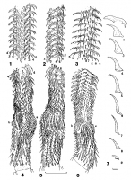
Figs. 17. Pintneriella musculicola Yamaguti, 1934. Tentacular armature. Fig. 1. Metabasal armature, internal surface of
tentacle. Fig. 2. Metabasal armature, antibothridial surface of tentacle. Fig.... MoreFigs. 17. Pintneriella musculicola Yamaguti, 1934. Tentacular armature. Fig. 1. Metabasal armature, internal surface of
tentacle. Fig. 2. Metabasal armature, antibothridial surface of tentacle. Fig. 3. Metabasal armature, external surface of tentacle.
Fig. 4. Basal armature and origin of metabasal armature, internal surface of tentacle. Fig. 5. Basal armature and origin of
metabasal armature, bothridial surface of tentacle. Fig. 6. Basal armature and origin of metabasal armature, external surface of
tentacle. Fig. 7. Profiles of hooks of principal row, 19. Abbreviations: a antibothridial, b bothridial. Scale bars: Figs. 16 =
0.1 mm; Fig. 7 = 0.01 mm. |
Line Drawing 2

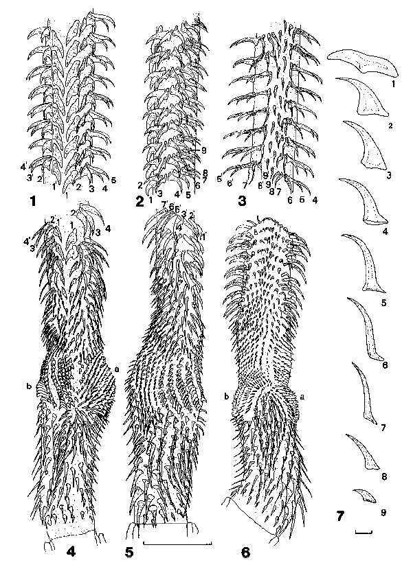
Figs. 811. Pintneriella musculicola Yamaguti, 1934. Fig. 8. Scolex. Fig. 9. Tentacular bulb. Fig. 10. Mature segment; vitelline
follicles shown on lateral margins of segment only. Fig. 11. Post-matu... MoreFigs. 811. Pintneriella musculicola Yamaguti, 1934. Fig. 8. Scolex. Fig. 9. Tentacular bulb. Fig. 10. Mature segment; vitelline
follicles shown on lateral margins of segment only. Fig. 11. Post-mature segment showing enlargement of uterus and patent
uterine pore. Abbreviations: u uterus, up uterine pore, vi vitelline follicles. Scale bars = 0.1 mm. Fig. 12. Vagina, uterus and uterine pore. For the sake of clarity, the
dorso-ventral relationships of the vagina and uterus have not been preserved. Fig. 13. Cirrus sac and seminal vesicles, lateral
view, cirrus inverted. Fig. 14. Cirrus sac and seminal vesicles, lateral view, cirrus everted. Fig. 15. Transverse section of mature
segment at level of ovary showing four-lobed ovary and single layer of testes. Fig. 16. Transverse histological section through
genital atrium and terminal genitalia showing vagina opening to atrium independently of cirrus sac and genital ducts passing
ventrally to osmoregulatory canals. Fig. 17. Gravid segment with grossly distended uterus filled with eggs. Abbreviations: ao
accessory osmoregulatory canal, do dorsal osmoregulatory canal, esv external seminal vesicle, isv internal seminal vesicle,
m Mehlis gland, o ovary, sr seminal receptacle, t testis, u uterus, up uterine pore, v vagina, vd vas deferens, vi
vitelline follicles, vo ventral osmoregulatory canal. Scale bars = 0.1 mm. |
Photo Micrograph
|
Scanning Electron Micrograph
|
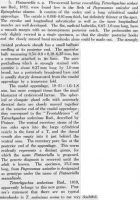


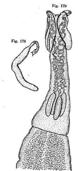 Fig. 178. Pintneriella musculicola. Type 25.2 mm long.
Fig. 179. Anterior portion of same.
Fig. 178. Pintneriella musculicola. Type 25.2 mm long.
Fig. 179. Anterior portion of same. 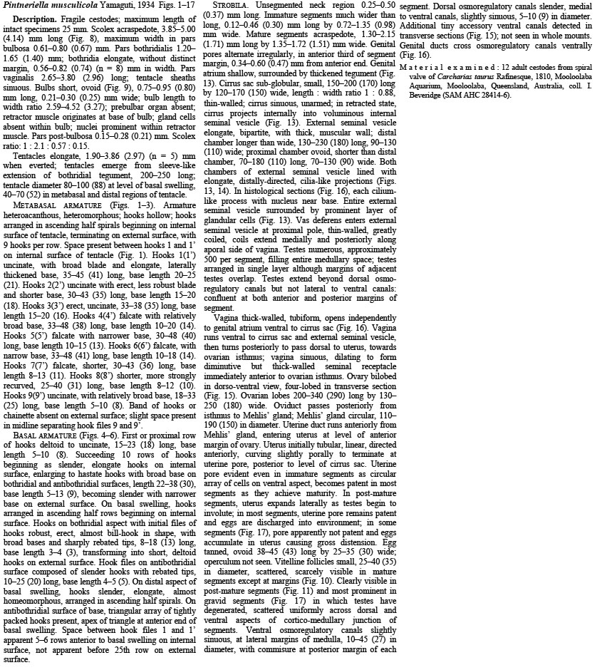
 Figs. 17. Pintneriella musculicola Yamaguti, 1934. Tentacular armature. Fig. 1. Metabasal armature, internal surface of
tentacle. Fig. 2. Metabasal armature, antibothridial surface of tentacle. Fig. 3. Metabasal armature, external surface of tentacle.
Fig. 4. Basal armature and origin of metabasal armature, internal surface of tentacle. Fig. 5. Basal armature and origin of
metabasal armature, bothridial surface of tentacle. Fig. 6. Basal armature and origin of metabasal armature, external surface of
tentacle. Fig. 7. Profiles of hooks of principal row, 19. Abbreviations: a antibothridial, b bothridial. Scale bars: Figs. 16 =
0.1 mm; Fig. 7 = 0.01 mm.
Figs. 17. Pintneriella musculicola Yamaguti, 1934. Tentacular armature. Fig. 1. Metabasal armature, internal surface of
tentacle. Fig. 2. Metabasal armature, antibothridial surface of tentacle. Fig. 3. Metabasal armature, external surface of tentacle.
Fig. 4. Basal armature and origin of metabasal armature, internal surface of tentacle. Fig. 5. Basal armature and origin of
metabasal armature, bothridial surface of tentacle. Fig. 6. Basal armature and origin of metabasal armature, external surface of
tentacle. Fig. 7. Profiles of hooks of principal row, 19. Abbreviations: a antibothridial, b bothridial. Scale bars: Figs. 16 =
0.1 mm; Fig. 7 = 0.01 mm. 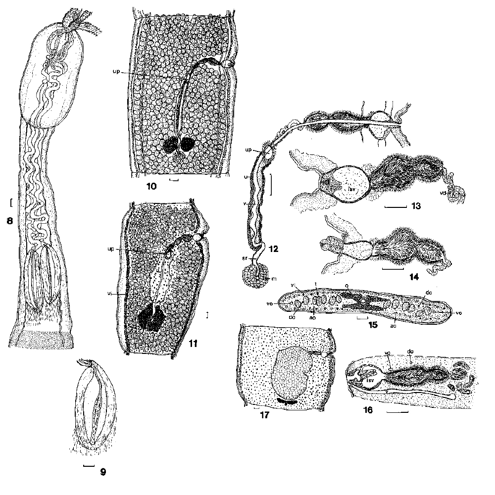 Figs. 811. Pintneriella musculicola Yamaguti, 1934. Fig. 8. Scolex. Fig. 9. Tentacular bulb. Fig. 10. Mature segment; vitelline
follicles shown on lateral margins of segment only. Fig. 11. Post-mature segment showing enlargement of uterus and patent
uterine pore. Abbreviations: u uterus, up uterine pore, vi vitelline follicles. Scale bars = 0.1 mm. Fig. 12. Vagina, uterus and uterine pore. For the sake of clarity, the
dorso-ventral relationships of the vagina and uterus have not been preserved. Fig. 13. Cirrus sac and seminal vesicles, lateral
view, cirrus inverted. Fig. 14. Cirrus sac and seminal vesicles, lateral view, cirrus everted. Fig. 15. Transverse section of mature
segment at level of ovary showing four-lobed ovary and single layer of testes. Fig. 16. Transverse histological section through
genital atrium and terminal genitalia showing vagina opening to atrium independently of cirrus sac and genital ducts passing
ventrally to osmoregulatory canals. Fig. 17. Gravid segment with grossly distended uterus filled with eggs. Abbreviations: ao
accessory osmoregulatory canal, do dorsal osmoregulatory canal, esv external seminal vesicle, isv internal seminal vesicle,
m Mehlis gland, o ovary, sr seminal receptacle, t testis, u uterus, up uterine pore, v vagina, vd vas deferens, vi
vitelline follicles, vo ventral osmoregulatory canal. Scale bars = 0.1 mm.
Figs. 811. Pintneriella musculicola Yamaguti, 1934. Fig. 8. Scolex. Fig. 9. Tentacular bulb. Fig. 10. Mature segment; vitelline
follicles shown on lateral margins of segment only. Fig. 11. Post-mature segment showing enlargement of uterus and patent
uterine pore. Abbreviations: u uterus, up uterine pore, vi vitelline follicles. Scale bars = 0.1 mm. Fig. 12. Vagina, uterus and uterine pore. For the sake of clarity, the
dorso-ventral relationships of the vagina and uterus have not been preserved. Fig. 13. Cirrus sac and seminal vesicles, lateral
view, cirrus inverted. Fig. 14. Cirrus sac and seminal vesicles, lateral view, cirrus everted. Fig. 15. Transverse section of mature
segment at level of ovary showing four-lobed ovary and single layer of testes. Fig. 16. Transverse histological section through
genital atrium and terminal genitalia showing vagina opening to atrium independently of cirrus sac and genital ducts passing
ventrally to osmoregulatory canals. Fig. 17. Gravid segment with grossly distended uterus filled with eggs. Abbreviations: ao
accessory osmoregulatory canal, do dorsal osmoregulatory canal, esv external seminal vesicle, isv internal seminal vesicle,
m Mehlis gland, o ovary, sr seminal receptacle, t testis, u uterus, up uterine pore, v vagina, vd vas deferens, vi
vitelline follicles, vo ventral osmoregulatory canal. Scale bars = 0.1 mm. 

