Line Drawing 1
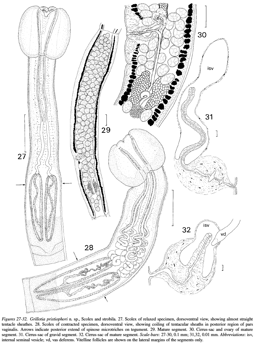
Figures 27-32. Grillotia pristiophori n. sp., Scolex and strobila. 27. Scolex of relaxed specimen, dorsoventral view, showing almost straight
tentacle sheathes. 28. Scolex of contracted specimen, dor... MoreFigures 27-32. Grillotia pristiophori n. sp., Scolex and strobila. 27. Scolex of relaxed specimen, dorsoventral view, showing almost straight
tentacle sheathes. 28. Scolex of contracted specimen, dorsoventral view, showing coiling of tentacular sheaths in posterior region of pars
vaginalis. Arrows indicate posterior extend of spinose microtriches on tegument. 29. Mature segment. 30. Cirrus-sac and ovary of mature
segment. 31. Cirrus-sac of gravid segment. 32. Cirrus-sac of mature segment. Scale-bars: 27-30, 0.1 mm; 31,32, 0.01 mm. Abbreviations: isv,
internal seminal vesicle; vd, vas deferens. Vitelline follicles are shown on the lateral margins of the segments only. |
Line Drawing 2
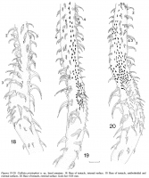
Figures 18-20. Grillotia pristiophori n. sp., basal armature. 18. Base of tentacle, internal surface. 19. Base of tentacle, antibothridial and
external surfaces. 20. Base of tentacle, external surfac... MoreFigures 18-20. Grillotia pristiophori n. sp., basal armature. 18. Base of tentacle, internal surface. 19. Base of tentacle, antibothridial and
external surfaces. 20. Base of tentacle, external surface. Scale-bar: 0.01 mm. |
Photo Micrograph

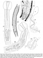
Figures 21-26. Grillotia pristiophori n. sp., tentacular armature. 21.Metabasal region, internal surface. 22.Metabasal region, bothridial surface.
23. Metabasal region, external surface. 24. Hook pro... MoreFigures 21-26. Grillotia pristiophori n. sp., tentacular armature. 21.Metabasal region, internal surface. 22.Metabasal region, bothridial surface.
23. Metabasal region, external surface. 24. Hook profiles. 25. Hooks of files 1 and 1! from basal region showing differences in size and shape.
26. Schematic representation of pattern of metabasal tentacular armature in which the band of hooklets is reduced to a single file indicated here
as hooks 7(7!). Scale-bars: 0.01 mm. |
Scanning Electron Micrograph

Figures 33-36. Grillotia pristiophori n. sp., scanning electron micrographs. 33. Bothridium. 34. Distal tip of tentacle, external surface showing
single file of hooks in middle of external surface. 3... MoreFigures 33-36. Grillotia pristiophori n. sp., scanning electron micrographs. 33. Bothridium. 34. Distal tip of tentacle, external surface showing
single file of hooks in middle of external surface. 35. Scolex, showing spiniform microtriches restricted to anterior part of pars pedunculus
scolecis. 36. Basal armature of tentacle, external surface showing transformation of band of hooklets to two files anteriorly. Scale-bars: 33,35, 0.1 mm; 34,36,0.01 mm. |

 Figures 27-32. Grillotia pristiophori n. sp., Scolex and strobila. 27. Scolex of relaxed specimen, dorsoventral view, showing almost straight
tentacle sheathes. 28. Scolex of contracted specimen, dorsoventral view, showing coiling of tentacular sheaths in posterior region of pars
vaginalis. Arrows indicate posterior extend of spinose microtriches on tegument. 29. Mature segment. 30. Cirrus-sac and ovary of mature
segment. 31. Cirrus-sac of gravid segment. 32. Cirrus-sac of mature segment. Scale-bars: 27-30, 0.1 mm; 31,32, 0.01 mm. Abbreviations: isv,
internal seminal vesicle; vd, vas deferens. Vitelline follicles are shown on the lateral margins of the segments only.
Figures 27-32. Grillotia pristiophori n. sp., Scolex and strobila. 27. Scolex of relaxed specimen, dorsoventral view, showing almost straight
tentacle sheathes. 28. Scolex of contracted specimen, dorsoventral view, showing coiling of tentacular sheaths in posterior region of pars
vaginalis. Arrows indicate posterior extend of spinose microtriches on tegument. 29. Mature segment. 30. Cirrus-sac and ovary of mature
segment. 31. Cirrus-sac of gravid segment. 32. Cirrus-sac of mature segment. Scale-bars: 27-30, 0.1 mm; 31,32, 0.01 mm. Abbreviations: isv,
internal seminal vesicle; vd, vas deferens. Vitelline follicles are shown on the lateral margins of the segments only.  Figures 18-20. Grillotia pristiophori n. sp., basal armature. 18. Base of tentacle, internal surface. 19. Base of tentacle, antibothridial and
external surfaces. 20. Base of tentacle, external surface. Scale-bar: 0.01 mm.
Figures 18-20. Grillotia pristiophori n. sp., basal armature. 18. Base of tentacle, internal surface. 19. Base of tentacle, antibothridial and
external surfaces. 20. Base of tentacle, external surface. Scale-bar: 0.01 mm. 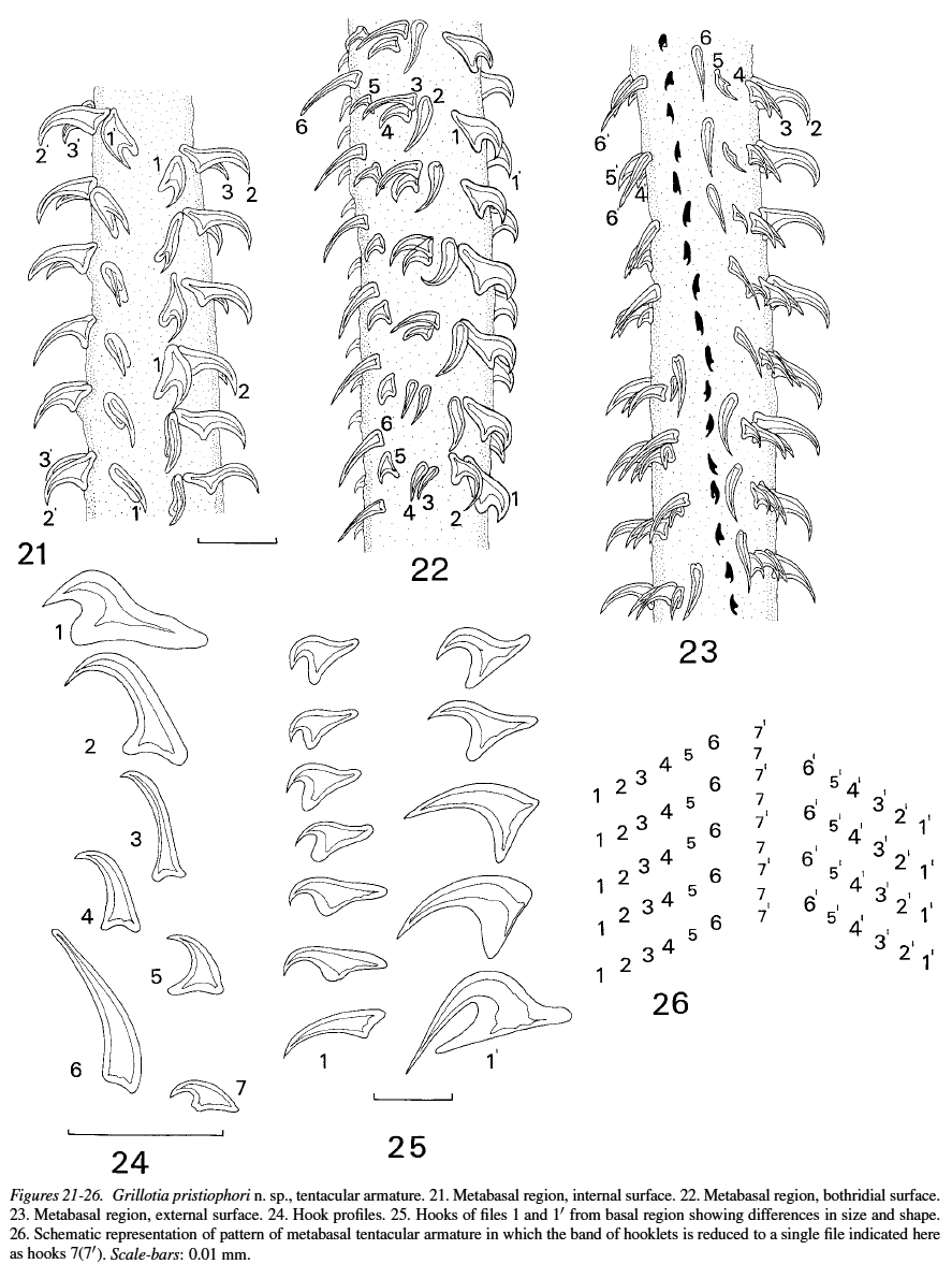 Figures 21-26. Grillotia pristiophori n. sp., tentacular armature. 21.Metabasal region, internal surface. 22.Metabasal region, bothridial surface.
23. Metabasal region, external surface. 24. Hook profiles. 25. Hooks of files 1 and 1! from basal region showing differences in size and shape.
26. Schematic representation of pattern of metabasal tentacular armature in which the band of hooklets is reduced to a single file indicated here
as hooks 7(7!). Scale-bars: 0.01 mm.
Figures 21-26. Grillotia pristiophori n. sp., tentacular armature. 21.Metabasal region, internal surface. 22.Metabasal region, bothridial surface.
23. Metabasal region, external surface. 24. Hook profiles. 25. Hooks of files 1 and 1! from basal region showing differences in size and shape.
26. Schematic representation of pattern of metabasal tentacular armature in which the band of hooklets is reduced to a single file indicated here
as hooks 7(7!). Scale-bars: 0.01 mm. 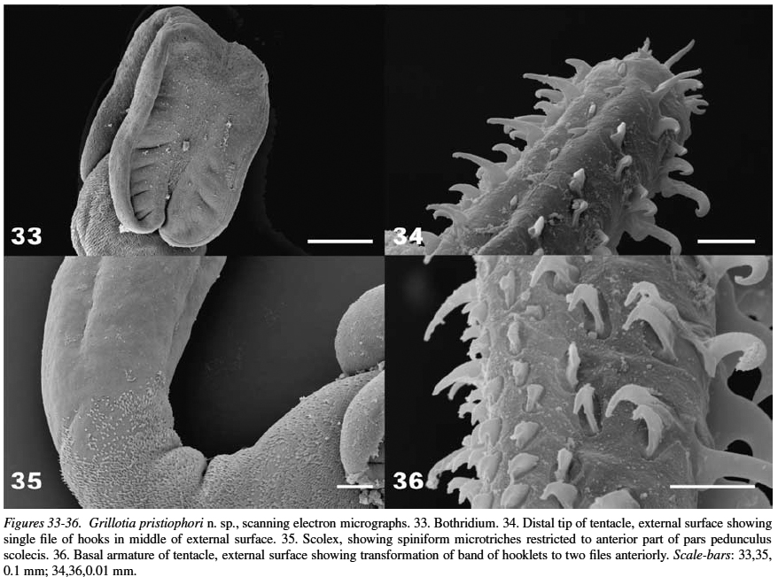 Figures 33-36. Grillotia pristiophori n. sp., scanning electron micrographs. 33. Bothridium. 34. Distal tip of tentacle, external surface showing
single file of hooks in middle of external surface. 35. Scolex, showing spiniform microtriches restricted to anterior part of pars pedunculus
scolecis. 36. Basal armature of tentacle, external surface showing transformation of band of hooklets to two files anteriorly. Scale-bars: 33,35, 0.1 mm; 34,36,0.01 mm.
Figures 33-36. Grillotia pristiophori n. sp., scanning electron micrographs. 33. Bothridium. 34. Distal tip of tentacle, external surface showing
single file of hooks in middle of external surface. 35. Scolex, showing spiniform microtriches restricted to anterior part of pars pedunculus
scolecis. 36. Basal armature of tentacle, external surface showing transformation of band of hooklets to two files anteriorly. Scale-bars: 33,35, 0.1 mm; 34,36,0.01 mm. 

