Line Drawing 1
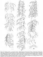
Figures 1-10. Grillotia australis n. sp., tentacular armature. 1. Metabasal region, internal surface of tentacle. 2. Distal region of tentacle, bothridial
surface. Note differences in shape of hooks ... MoreFigures 1-10. Grillotia australis n. sp., tentacular armature. 1. Metabasal region, internal surface of tentacle. 2. Distal region of tentacle, bothridial
surface. Note differences in shape of hooks 1(1!) when compared with Figure 1. 3. Distal region, external surface of tentacle. 4.Metabasal
region, bothridial surface of tentacle. 5. Basal region of tentacle, internal surface. 6. Basal region of tentacle, bothridial surface. 7. Basal region
of tentacle, antibothridial surface. 8. Basal region of tentacle, external surface. 9. Profiles of hooks 1 to 4 in metabasal region. 10. Schematic
representation of pattern of metabasal tentacular armature. Scale-bar: 0.01 mm. Hooks of principal rows are numbered, intercalary rows are
designated by letters and hooklets of the external surface are indicated in solid black. |
Line Drawing 2
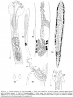
Figures 11-17. Grillotia australis n. sp., scolex and strobila. 11. Scolex, dorso-ventral view. 12. Gravid segment. 13. Mature segment, genital
ducts. 14. Mature segment. 15. Eggs. 16. Terminal genit... MoreFigures 11-17. Grillotia australis n. sp., scolex and strobila. 11. Scolex, dorso-ventral view. 12. Gravid segment. 13. Mature segment, genital
ducts. 14. Mature segment. 15. Eggs. 16. Terminal genitalia. 17. Scolex, lateral view. Scale-bars: 11-14,16,17, 0.1 mm; 15, 0.01 mm.
Abbreviations: asv, accessory seminal vesicle; esv, external seminal vesicle, isv, internal seminal vesicle; v, vagina; vd, vas deferens. Vitelline
follicles are shown on the lateral margins of the segments only. |
Photo Micrograph
|
Scanning Electron Micrograph
|


 Figures 1-10. Grillotia australis n. sp., tentacular armature. 1. Metabasal region, internal surface of tentacle. 2. Distal region of tentacle, bothridial
surface. Note differences in shape of hooks 1(1!) when compared with Figure 1. 3. Distal region, external surface of tentacle. 4.Metabasal
region, bothridial surface of tentacle. 5. Basal region of tentacle, internal surface. 6. Basal region of tentacle, bothridial surface. 7. Basal region
of tentacle, antibothridial surface. 8. Basal region of tentacle, external surface. 9. Profiles of hooks 1 to 4 in metabasal region. 10. Schematic
representation of pattern of metabasal tentacular armature. Scale-bar: 0.01 mm. Hooks of principal rows are numbered, intercalary rows are
designated by letters and hooklets of the external surface are indicated in solid black.
Figures 1-10. Grillotia australis n. sp., tentacular armature. 1. Metabasal region, internal surface of tentacle. 2. Distal region of tentacle, bothridial
surface. Note differences in shape of hooks 1(1!) when compared with Figure 1. 3. Distal region, external surface of tentacle. 4.Metabasal
region, bothridial surface of tentacle. 5. Basal region of tentacle, internal surface. 6. Basal region of tentacle, bothridial surface. 7. Basal region
of tentacle, antibothridial surface. 8. Basal region of tentacle, external surface. 9. Profiles of hooks 1 to 4 in metabasal region. 10. Schematic
representation of pattern of metabasal tentacular armature. Scale-bar: 0.01 mm. Hooks of principal rows are numbered, intercalary rows are
designated by letters and hooklets of the external surface are indicated in solid black.  Figures 11-17. Grillotia australis n. sp., scolex and strobila. 11. Scolex, dorso-ventral view. 12. Gravid segment. 13. Mature segment, genital
ducts. 14. Mature segment. 15. Eggs. 16. Terminal genitalia. 17. Scolex, lateral view. Scale-bars: 11-14,16,17, 0.1 mm; 15, 0.01 mm.
Abbreviations: asv, accessory seminal vesicle; esv, external seminal vesicle, isv, internal seminal vesicle; v, vagina; vd, vas deferens. Vitelline
follicles are shown on the lateral margins of the segments only.
Figures 11-17. Grillotia australis n. sp., scolex and strobila. 11. Scolex, dorso-ventral view. 12. Gravid segment. 13. Mature segment, genital
ducts. 14. Mature segment. 15. Eggs. 16. Terminal genitalia. 17. Scolex, lateral view. Scale-bars: 11-14,16,17, 0.1 mm; 15, 0.01 mm.
Abbreviations: asv, accessory seminal vesicle; esv, external seminal vesicle, isv, internal seminal vesicle; v, vagina; vd, vas deferens. Vitelline
follicles are shown on the lateral margins of the segments only. 

