Cestode Scientific Name
| Species ID | 4751 |
|---|---|
| Order | Trypanorhyncha |
| Family | Pseudotobothriidae |
| Subfamily | |
| Genus | Pseudotobothrium |
| Species | dipsacum |
| Authority | (Linton, 1897) Dollfus, 1942 |
| Taxonomic Status | Valid |
| Valid Name | |
| Synonyms | Otobothrium dipsacum Linton, 1897 |
| Genus Record | No |
| Type Species | Yes |
| Verified | Yes |
| Verified By | I. Beveridge |
| Citation(s) |
Linton, E. 1897. Notes on cestode parasites of fishes. Proceedings of the United States National Museum 20: 423-456. (272) Download PDFDollfus, R.-P. 1942. Études critiques sur les tétrarhynques du Muséum de Paris. Archives du Muséum National dHistoire Naturelle, 6. Série 19: 7-466. (170) Download PDF |
| Redescription |
Beveridge, I., R. A. Campbell, and M. K. Jones. 2000. New records of the cestode genus Pseudotobothrium (Trypnaorhyncha: Otobothriidae) from Australian fishes. Transactions of the Royal Society of South Australia 124(2): 151-162. (4317) Download PDF |
| Scientific Name Notes |
Record Data
| Date (MM/DD/YYYY) | Action | User Name |
|---|---|---|
| 11/30/-0001 | Created | I. Beveridge |
| 05/07/2015 | Modified | |
| 06/02/2015 | Modified | I. Beveridge |
| 05/13/2016 | Modified | B. Barbeau |
| 08/18/2017 | Modified | K. Herzog |
Type Host
| Host Class | Actinopterygii | ||||||
|---|---|---|---|---|---|---|---|
| Host Order | Perciformes | ||||||
| Host Family | Pomatomidae | ||||||
|
Type Host (Literal) |
|
||||||
|
Type Host (Valid) |
|
||||||
| Additional Host(s) | |||||||
| Site in Host | viscera | ||||||
| Host Notes | Numerous teleosts act as additional hosts: see Beveridge et al. (2000); Palm (2004) |
Type Locality
| Country | U.S.A. |
|---|---|
| Body of Water | Atlantic Ocean |
| Island(s) | |
| City/Region | New England |
| Coordinates | |
| DD Latitude | |
| DD Longitude | |
| Additional Localities | |
| Locality Notes | Cosmopolitan; see Beveridge et al. (2000); Palm (2004) |
Specimens
| Type Material | USNPC 4794 (holotype) |
|---|---|
| Total Number of Type Specimens | |
| Voucher Material | |
| Specimen Notes |
Data are given as in original description unless otherwise indicated.

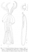
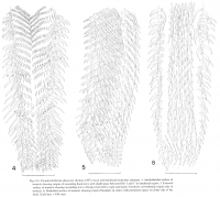
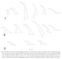

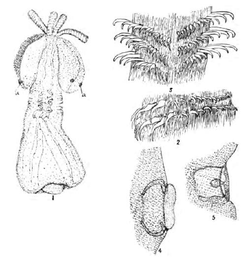 Plate VI. Otobothrium dipsacum, new species, from Pomatomus saltatrix. 1. Embryo removed from blastocyst. a, ciliated organs of bothria (shown enlarged in figs. 4,5). Enlarged twenty-seven times. 2. Two transverse rows of hooks on proboscis. Enlarged three hundred times. 3. Obverse side of proboscis from that shown in fig. 2. 4. Ciliated organs (rudimentary sense organs) of bothria. Enlarged two hundred and twenty-five times. 5. The same invaginated.
 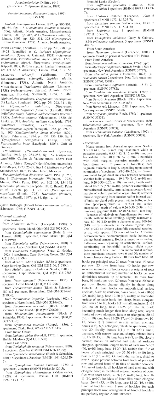 Beveridge et al. (2000), pg. 152
 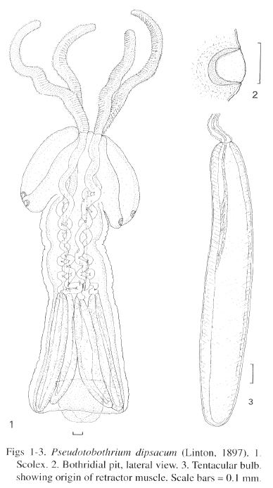 Figs 1-3. Pseudobothrium dipsacwn (Linton, 1897). I Scolex. 2. Bothridial pit, lateral view. 3. Tentacular bulb showing origin of retractor muscle. Scale bars= 0.1 mm.
  Figs 4-6 . Pseudotobothrium dipsacum (Linton, 1897) , basal and metabasal tentacular armature. 4. Antibothridial surface of tentacle showing origins of ascending hook rows with slight space between files I and I' in metabasal region. 5. External surface of tentacle showing ascending rows of hooks from left to right and band of hook lets on bothridial (right) side of tentacle. 6. Bothridial surface of tentacle showing band of hooklets in centre with prominent space on either side of the band. Scale bars = 0.01 mm.
 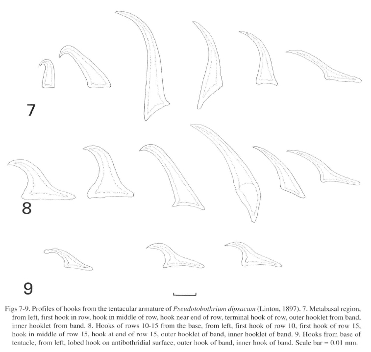 Figs 7-9. Profiles of hooks from the tentacular armature of Pseudotobothrium dipsacum (Linton , 1897). 7. Metabasal region,
from left , fir st hook in row , hook in middle of row, hook near end of row, terminal hook of row, outer hook let from band, inner hooklet from band. 8. Hook s of rows 10- 15 from the base, from left, fir st hook or row 10. first hook of row 15, hook in middle of row 15, hook at end of row 15, outer booklet of band, inner hooklet of band. 9. Hooks from base of tentacle, from left, lobed hook on antibothridial surface, outer hook of band, inner hook of band. Scale bar= 0.01 mm.
Figs 7-9. Profiles of hooks from the tentacular armature of Pseudotobothrium dipsacum (Linton , 1897). 7. Metabasal region,
from left , fir st hook in row , hook in middle of row, hook near end of row, terminal hook of row, outer hook let from band, inner hooklet from band. 8. Hook s of rows 10- 15 from the base, from left, fir st hook or row 10. first hook of row 15, hook in middle of row 15, hook at end of row 15, outer booklet of band, inner hooklet of band. 9. Hooks from base of tentacle, from left, lobed hook on antibothridial surface, outer hook of band, inner hook of band. Scale bar= 0.01 mm.  Dollfus (1942), pg. 253
 Best viewed in Firefox


