Line Drawing 1

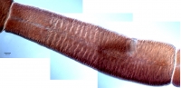
Figure 2 Line drawings of Rhinoptericola megacantha Carvajal & Campbell, 1975. (A) Whole worm (USNM 1661579; voucher). (B) Scolex (USNM 1661577; voucher). (C) Terminal proglottid (USNM 1661584; vouche... MoreFigure 2 Line drawings of Rhinoptericola megacantha Carvajal & Campbell, 1975. (A) Whole worm (USNM 1661579; voucher). (B) Scolex (USNM 1661577; voucher). (C) Terminal proglottid (USNM 1661584; voucher); circumcortical vitelline follicles are drawn only on the lateral margins and in the region delimited by dashed lines. Arrowheads indicate the level at which the sections in Fig. 5 were taken. |
Line Drawing 2
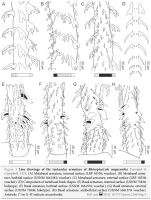
Figure 3 Line drawings of the tentacular armature of Rhinoptericola megacantha Carvajal & Campbell, 1975. (A) Metabasal armature, internal surface (LRP 10538; voucher). (B) Metabasal armature, bothria... MoreFigure 3 Line drawings of the tentacular armature of Rhinoptericola megacantha Carvajal & Campbell, 1975. (A) Metabasal armature, internal surface (LRP 10538; voucher). (B) Metabasal armature, bothrial surface (USNM 1661582; voucher). (C) Metabasal armature, external surface (LRP 10538; voucher). (D) Comparison of metabasal hook shapes. (E) Basal armature, internal surface (USNM 73836; holotype). (F) Basal armature, bothrial surface (USNM 1661576; voucher). (G) Basal armature, external surface (USNM 73836; holotype). (H) Basal armature, antibothrial surface (USNM 1661579; voucher). Asterisks (*) in EH indicate macrohooks. |
Photo Micrograph
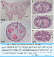
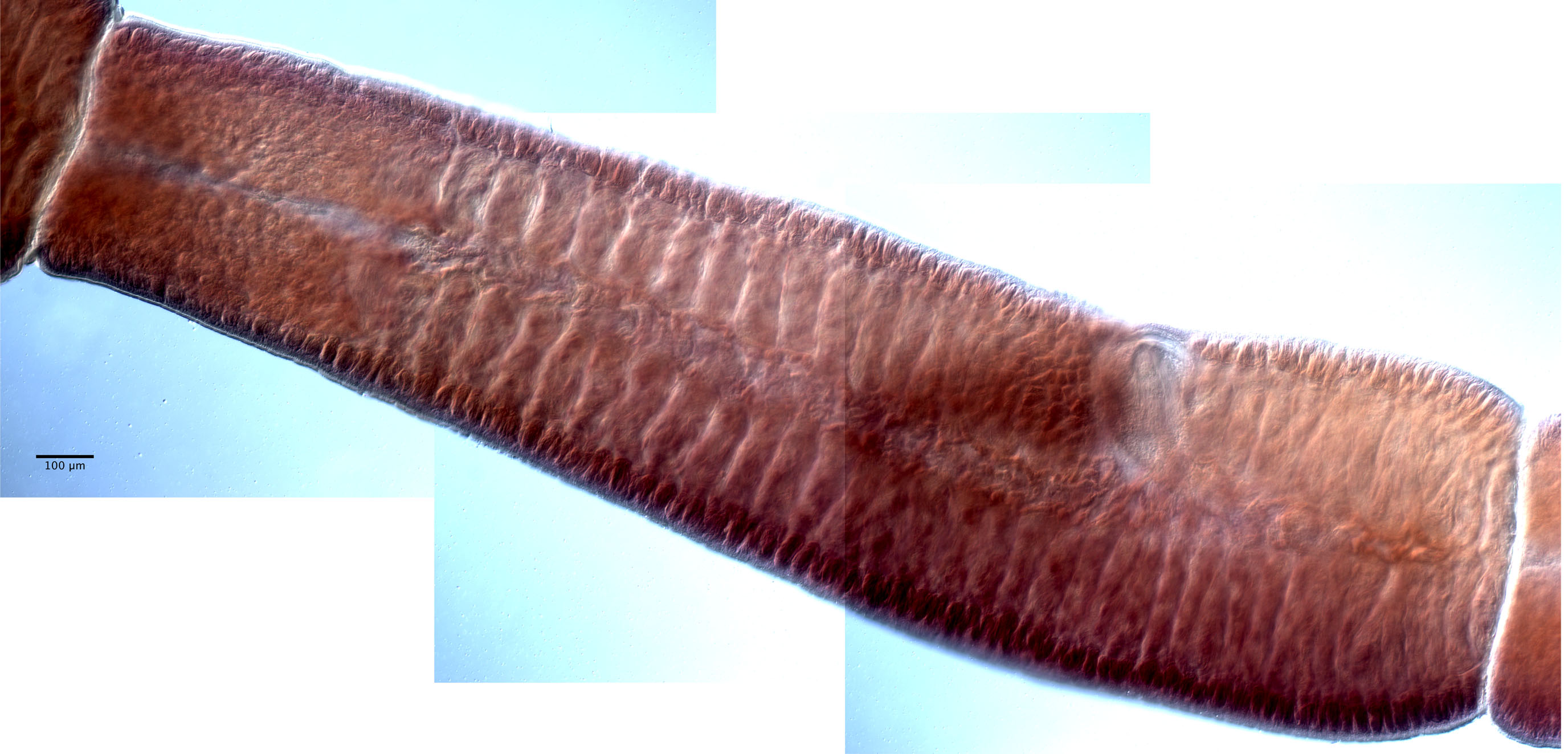
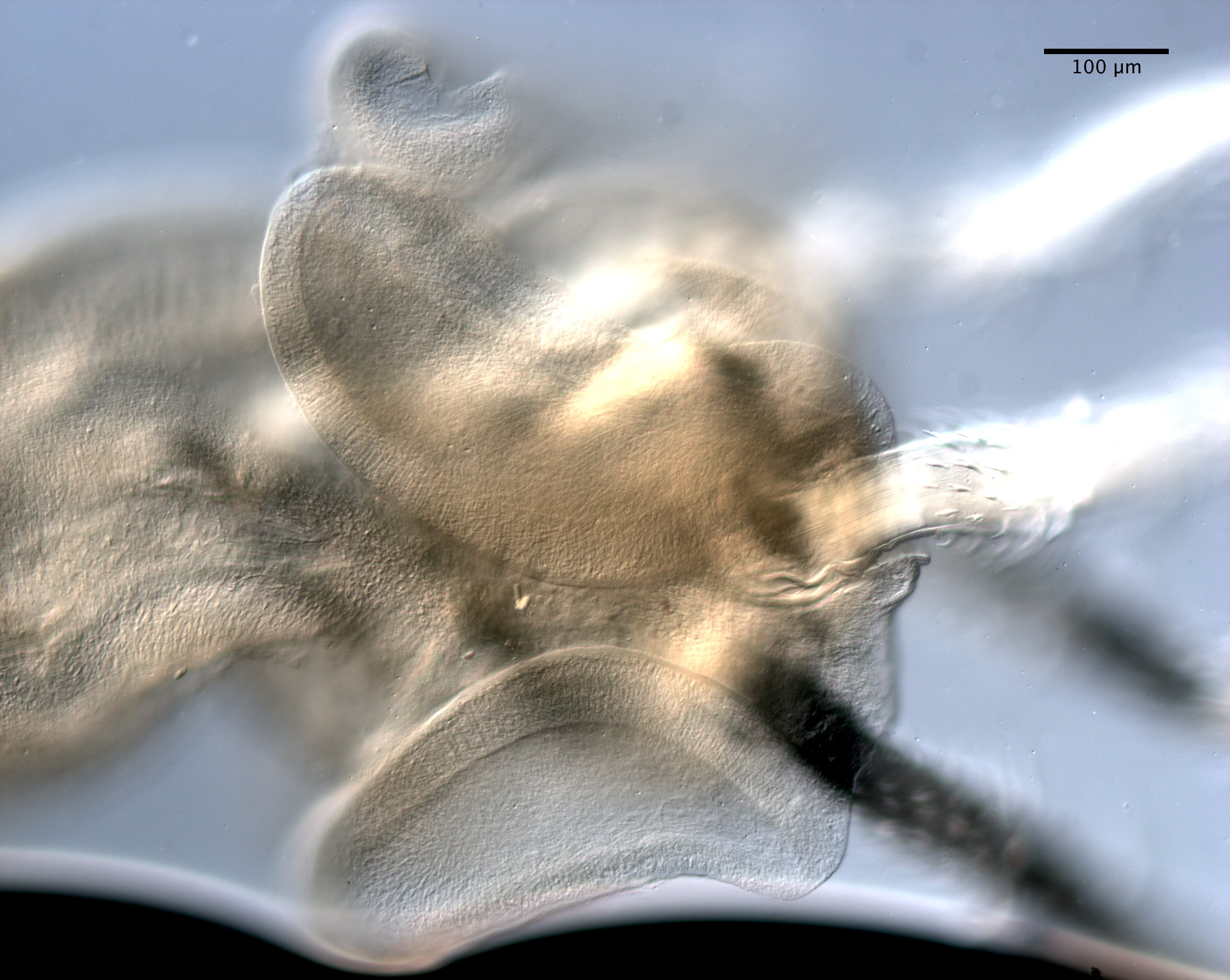
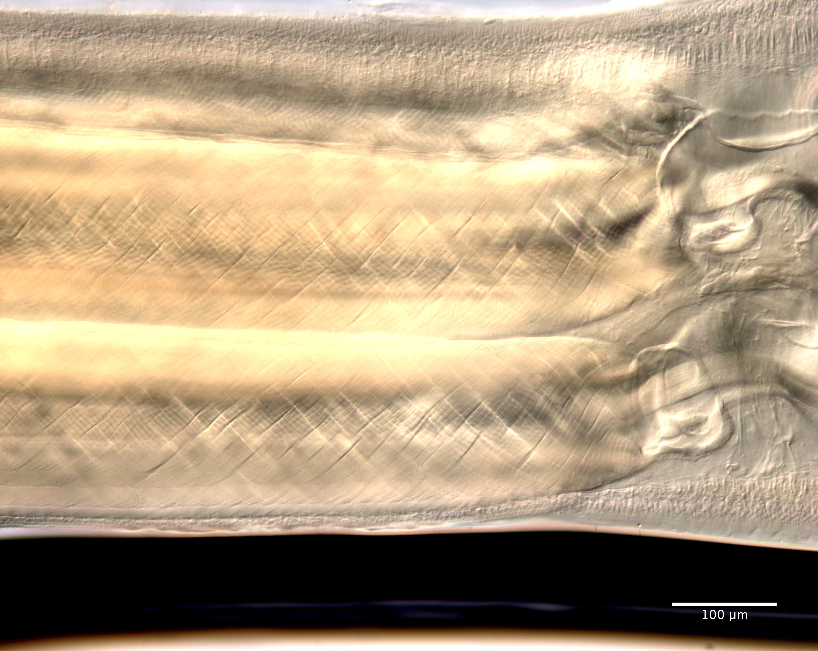
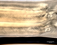
Figure 5 Light micrographs of cross-sections of Rhinoptericola megacantha Carvajal & Campbell, 1975. (A) Scolex at the level of the bothria. (B) Scolex at the level of the prebulbar organs. (C) Mature... MoreFigure 5 Light micrographs of cross-sections of Rhinoptericola megacantha Carvajal & Campbell, 1975. (A) Scolex at the level of the bothria. (B) Scolex at the level of the prebulbar organs. (C) Mature proglottid anterior to the genital pores. (D) Mature proglottid at the anterior margin of the ovary. (E) Mature proglottid at the level of the Mehlis gland. Abbreviations: BO, bothrium; BU, bulb; DEV, dorsal excretory vessel; M, Mehlis gland; O, ovary; PBO, prebulbar organ; RM, retractor muscle; T, testis; TE, tentacle; TS, tentacle sheath; U, uterus; UD, uterine duct; UTD, uterine diverticulum; VA, vagina; VEV, ventral excretory vessel; VID, vitelline duct; VF, vitelline follicle |
Scanning Electron Micrograph

Figure 4 Scanning electron micrographs of Rhinoptericola megacantha Carvajal & Campbell, 1975. (A) Scolex; small letters indicate the location of details shown in (IK). (B) Bothria and tentacular arm... MoreFigure 4 Scanning electron micrographs of Rhinoptericola megacantha Carvajal & Campbell, 1975. (A) Scolex; small letters indicate the location of details shown in (IK). (B) Bothria and tentacular armature; small letters indicate the location of details shown in (DH). (C) Surface of everted cirrus. (D) Distal bothrial surface. (E) Proximal bothria surface near the bothrial rim. (F) Bothrial surface away from the bothrial rim. (G) Surface of the scolex proper between the bothria. (H) Surface of the scolex proper at the apex. (I) Surface of the pars vaginalis. (J) Surface of the pars bulbosa. (K) Strobilar surface. (L) Separate male and female genital pores. (M) Metabasal armature, internal surface. (N) Metabasal armature, external surface. (O) Basal armature, internal surface. (P) Basal armature, bothrial surface. Asterisks (*) in O and P indicate macrohooks. |


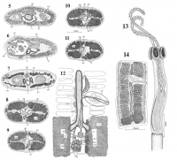


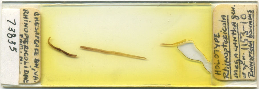
 FIGURES 1-4. Rhinoptericola megacantha. 1. Metabasal armature, internal face. 2. Basal armature, external face. 3. Basal armature, internal face. 4. Metabasal armature, external face. All figures to same scale.
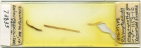
  FIGURES 5-12. Reproductive system of R. megacantha. Figs. 5-11. Transverse sections of semigravid segment. All drawn to same scale. 5. Anterior to cirrus pouch. 6. At level of genital pore. 7. Through proximal end of uterus. 8. Through lower half of ooreceptacle. 9-11. Consecutive sections showing origin of primary oviduct from ooreceptacle and junction with sperm duct from seminal receptacle. 12. Schematic diagram of reproductive system reconstructed with overlays from frontal and transverse serial sections. Abbreviations: CP, cirrus pouch; GP, genital pore; LD, uterine diverticulum; LM, longitudinal muscle; MG, Mehlis' gland; OC, oocapt; OD, primary oviduct; OO, ooreceptacle; ORC, ventral osmoregulatory canal; OV, ovary; SD, sperm duct; SO, secondary oviduct; SR, seminal recep- tacle; SV, seminal vesicle; T, testis; U, uterus; UD, uterine duct; UG, uterine glands; V, vagina; VD, vas deferens; VT, vitellaria; VTD, vitelline duct. FIGURES 13-14. Rhinoptericola megacantha. 13. Scolex (holotype). 14. Mature segment (holotype).
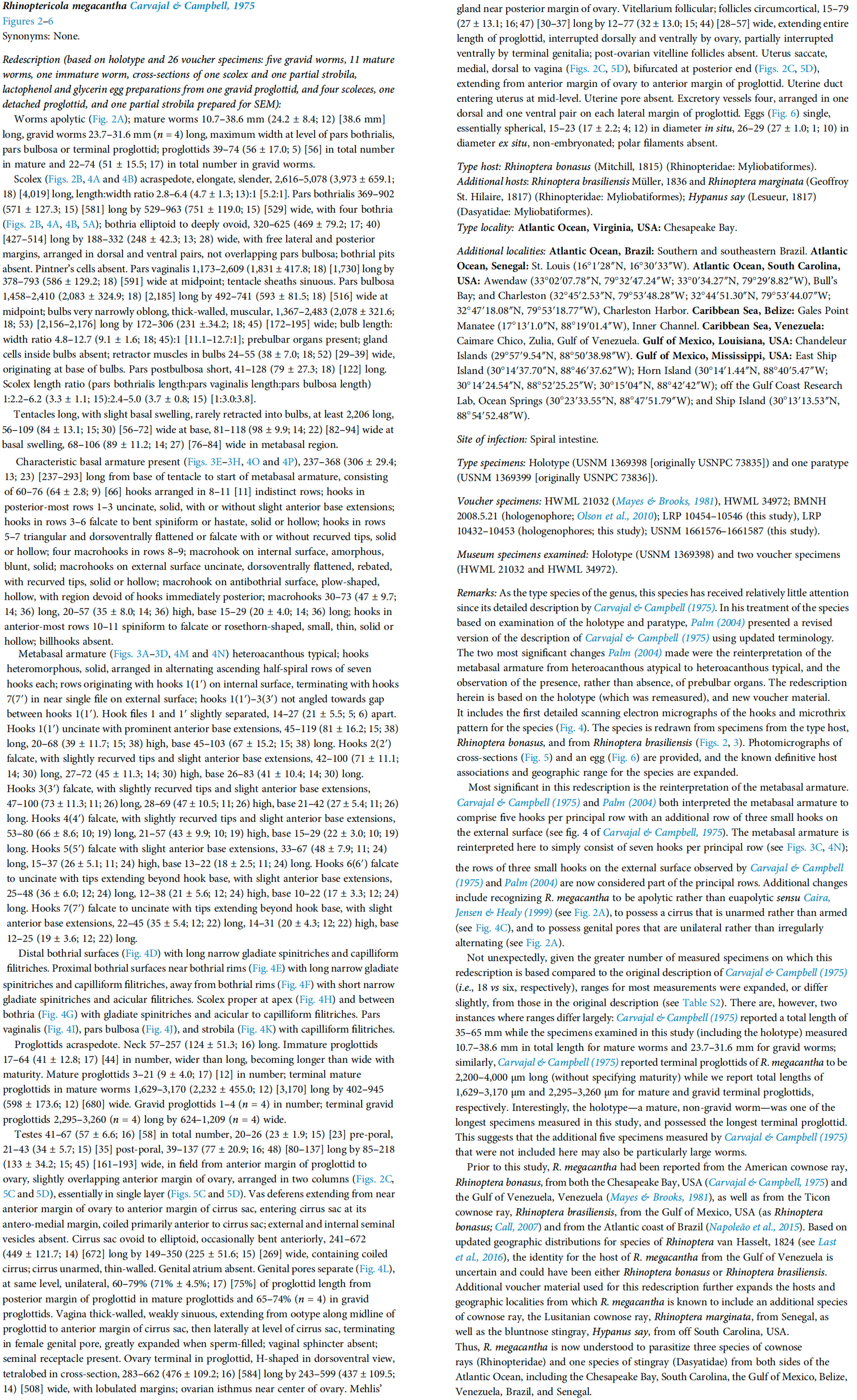
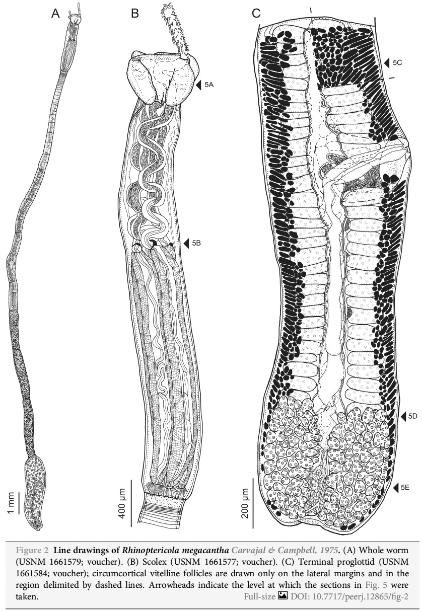 Figure 2 Line drawings of Rhinoptericola megacantha Carvajal & Campbell, 1975. (A) Whole worm (USNM 1661579; voucher). (B) Scolex (USNM 1661577; voucher). (C) Terminal proglottid (USNM 1661584; voucher); circumcortical vitelline follicles are drawn only on the lateral margins and in the region delimited by dashed lines. Arrowheads indicate the level at which the sections in Fig. 5 were taken.
 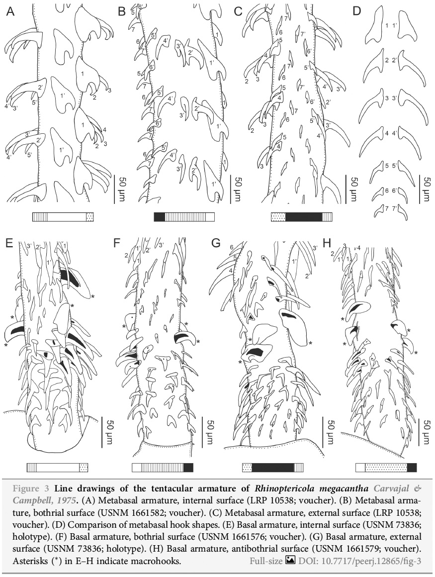 Figure 3 Line drawings of the tentacular armature of Rhinoptericola megacantha Carvajal & Campbell, 1975. (A) Metabasal armature, internal surface (LRP 10538; voucher). (B) Metabasal armature, bothrial surface (USNM 1661582; voucher). (C) Metabasal armature, external surface (LRP 10538; voucher). (D) Comparison of metabasal hook shapes. (E) Basal armature, internal surface (USNM 73836; holotype). (F) Basal armature, bothrial surface (USNM 1661576; voucher). (G) Basal armature, external surface (USNM 73836; holotype). (H) Basal armature, antibothrial surface (USNM 1661579; voucher). Asterisks (*) in EH indicate macrohooks.
  Figure 5 Light micrographs of cross-sections of Rhinoptericola megacantha Carvajal & Campbell, 1975. (A) Scolex at the level of the bothria. (B) Scolex at the level of the prebulbar organs. (C) Mature proglottid anterior to the genital pores. (D) Mature proglottid at the anterior margin of the ovary. (E) Mature proglottid at the level of the Mehlis gland. Abbreviations: BO, bothrium; BU, bulb; DEV, dorsal excretory vessel; M, Mehlis gland; O, ovary; PBO, prebulbar organ; RM, retractor muscle; T, testis; TE, tentacle; TS, tentacle sheath; U, uterus; UD, uterine duct; UTD, uterine diverticulum; VA, vagina; VEV, ventral excretory vessel; VID, vitelline duct; VF, vitelline follicle
  Figure 4 Scanning electron micrographs of Rhinoptericola megacantha Carvajal & Campbell, 1975. (A) Scolex; small letters indicate the location of details shown in (IK). (B) Bothria and tentacular armature; small letters indicate the location of details shown in (DH). (C) Surface of everted cirrus. (D) Distal bothrial surface. (E) Proximal bothria surface near the bothrial rim. (F) Bothrial surface away from the bothrial rim. (G) Surface of the scolex proper between the bothria. (H) Surface of the scolex proper at the apex. (I) Surface of the pars vaginalis. (J) Surface of the pars bulbosa. (K) Strobilar surface. (L) Separate male and female genital pores. (M) Metabasal armature, internal surface. (N) Metabasal armature, external surface. (O) Basal armature, internal surface. (P) Basal armature, bothrial surface. Asterisks (*) in O and P indicate macrohooks.